Иван Гайворонский - Anatomy of bone system. The manual for medical students / Анатомия костной системы. Учебное пособие для медицинских вузов
Здесь есть возможность читать онлайн «Иван Гайворонский - Anatomy of bone system. The manual for medical students / Анатомия костной системы. Учебное пособие для медицинских вузов» — ознакомительный отрывок электронной книги совершенно бесплатно, а после прочтения отрывка купить полную версию. В некоторых случаях можно слушать аудио, скачать через торрент в формате fb2 и присутствует краткое содержание. ISBN: , Жанр: Биология, Детская образовательная литература, foreign_language, на английском языке. Описание произведения, (предисловие) а так же отзывы посетителей доступны на портале библиотеки ЛибКат.
- Название:Anatomy of bone system. The manual for medical students / Анатомия костной системы. Учебное пособие для медицинских вузов
- Автор:
- Жанр:
- Год:неизвестен
- ISBN:978-5-299-00639-1
- Рейтинг книги:4 / 5. Голосов: 1
-
Избранное:Добавить в избранное
- Отзывы:
-
Ваша оценка:
Anatomy of bone system. The manual for medical students / Анатомия костной системы. Учебное пособие для медицинских вузов: краткое содержание, описание и аннотация
Предлагаем к чтению аннотацию, описание, краткое содержание или предисловие (зависит от того, что написал сам автор книги «Anatomy of bone system. The manual for medical students / Анатомия костной системы. Учебное пособие для медицинских вузов»). Если вы не нашли необходимую информацию о книге — напишите в комментариях, мы постараемся отыскать её.
Структура пособия соответствует современным стандартам медицинского образования в России и важнейшим Европейским стандартам. Английская и латинская терминология приведены в соответствии с Международной анатомической номенклатурой.
Anatomy of bone system. The manual for medical students / Анатомия костной системы. Учебное пособие для медицинских вузов — читать онлайн ознакомительный отрывок
Ниже представлен текст книги, разбитый по страницам. Система сохранения места последней прочитанной страницы, позволяет с удобством читать онлайн бесплатно книгу «Anatomy of bone system. The manual for medical students / Анатомия костной системы. Учебное пособие для медицинских вузов», без необходимости каждый раз заново искать на чём Вы остановились. Поставьте закладку, и сможете в любой момент перейти на страницу, на которой закончили чтение.
Интервал:
Закладка:
The atlas has an anterior arch, arcus anterior atlantis , instead of the body. Its anterior surface has an anterior tubercle, tuberculum anterius; on its posterior surface there is a small articular facet, fovea dentis , with which the II cervical vertebra is joined.
The lateral masses of atlas, massae laterales atlantis, are located on the both sides. Each of them bears an ellipsoid, concave superior articular surface, facies articularis superioris , joining with the corresponding occipital condyle. The inferior articular surfaces, facies articulares inferiores , form round, slightly concave articulate areas connected with the II cervical vertebra.
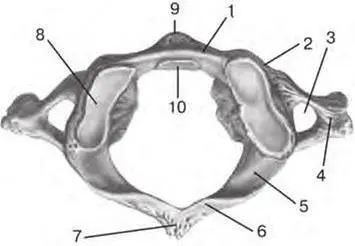
Fig. 2.3. I cervical vertebra (atlas) (superior aspect):
1 – anterior arch of atlas ( arcus anterior atlantis ); 2 – lateral mass ( massa lateralis ); 3 – transverse foramen ( foramen transversarium ); 4 – transverse process ( processus transversus ); 5 – groove for vertebral artery ( sulcus arteriae vertebralis ); 6 – posterior arch of atlas ( arcus posterior atlantis ); 7 – posterior tubercle ( tuberculum posterius ); 8 – superior articular surface ( facies articulars superior ); 9 – anterior tubercle ( tuberculum anterius ); 10 – facet for dens ( fovea dentis )
The posterior arch of the atlas, arcus posterior atlantis , corresponds to the arch of a typical vertebra. There is a reduced spinous process in the form of a small posterior tubercle, tuberculum posterius , on the back surface of the posterior arch. The groove for vertebral artery, sulcus arteriae vertebralis , passes on the superior surface of the posterior arch behind the lateral mass.
The vertebral foramen, foramen vertebrale , is bounded with arches and lateral masses. It is much larger than the vertebral foramina of other vertebrae, and only its posterior part corresponds to them. In its anterior part narrowed on the sides with the lateral masses, the dens of the II cervical vertebra is located. The transverse process, рrocessus transversus , is perforated by the transverse foramen, foramen transversarium , for the passage of blood vessels like transverse processes of the other cervical vertebrae. The ends of the transverse processes are slightly thickened. They have no anterior and posterior tubercles and no the groove for spinal nerve.
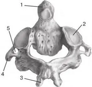
Fig. 2.4. II cervical vertebra (axis) (posterior aspect):
1 – dens ( dens ); 2 – superior articular facet ( facies articularis superioris ); 3 – spinous process ( processus spinosus ); 4 – transverse process ( processus transversus ); 5 – transverse foramen ( foramen transversarium )
The II cervical vertebra is termed the axis, axis (C II). Its distinctive feature is the presence of a dental process or dens, dens axis , projecting vertically from the superior surface of its body. The dens is the body of the atlas relocated during the development (fig. 2.4).
The dens plays the role of a pivot, around which the atlas together with the skull rotates to the right and to the left.
The dens of the II cervical vertebra is cylindrical, it has an apex, apex dentis , and articular facets on its anterior and posterior sides. The anterior articular facet, facies articularis anterior , articulates with fovea dentis of the anterior arch of the atlas; the posterior articular facet, facies articularis posterior , joins with the transverse ligament of the atlas. The axis has no superior articular processes. Instead of them there are slightly convex superior articular facets, facies articulares superiores , are on the sides of the dens. These facets articulate with the inferior articular surfaces on the lateral masses of the atlas. The ends of the transverse processes of the axis are slightly thickened like the ends of the I vertebra; there is the foramen transversarium at the base of each transverse process. The groove for spinal nerve is absent.
2.3. Thoracic Vertebrae
The thoracic vertebrae, vertebrae thoracicae (Th I– Th XII), are much bigger than the cervical vertebrae (fig. 2.1). The height and transverse size of the thoracic vertebrae bodies gradually increase from the I to XII vertebra. The distinctive feature of the thoracic vertebrae is the presence of the articular facets or demi-facets for articulation with ribs. These facets are located on the lateral sides of the bodies and on the transverse processes.
Most vertebrae have two demi-facets. They are directly in front of the pedicle of the vertebral arch: one – at superior edge, the other – at inferior edge. They are called the superior costal facet and inferior costal facet, respectively, fovea costalis superior et fovea costalis inferior . Each such facet (demi-facet to be more precise) of one vertebra, together with the nearest demi-facet of the adjacent vertebra, form an articular area for articulation with the head of the rib. An exception is the I vertebra: it has a complete facet to articulate with the I rib, and a demi-facet to articulate with the II rib. The X vertebra has only the superior demi-facet for articulation with the X rib. The XI and XII vertebrae have one complete facet for articulation with the corresponding ribs.
The articular processes of the thoracic vertebrae are located in the frontal plane. The articular surfaces of superior articular processes are directed backwards, the articular surfaces of the inferior articular processes are directed forwards. The transverse processes are directed laterally and backwards; their length increases from the I to the IX vertebra but then they become shorter. The ends of the transverse processes are thickened. They have a transverse costal facet, fovea costalis processus transversi , for articulation with the tubercle of the rib. The XI and XII vertebrae do not have such facet.
The spinous processes of the thoracic vertebrae are longer than the spinous processes of the cervical vertebrae; they are tilted downwards and lie on each other like tiles.
2.4. Lumbar Vertebrae
The distinctive feature of the lumbar vertebra, vertebrae lumbales (L I– L V), is a beanshaped massive body. The transverse size of the body is wider than the anteroposterior size. The height and width of the body gradually increases from the I to the V vertebrae. The vertebral foramen is large and triangular with rounded angles. The articular processes are well developed, their articular surfaces are located in the sagittal plane: the articular surfaces of the superior processes are directed medially, the articular surfaces of the inferior processes are directed laterally (fig. 2.5).
The superior articular process may have a rudimental tubercle named mammillary process, processus mamillaris . The lumbar vertebra`s transverse processes are formed by fusion of rudimental ribs with the transverse processes of the vertebra. They are located in the frontal plane, their ends are tilted backwards. Often the accessory process, processus accessorius , may be present at the base of the transverse process. The spinous processes are short and flat, their ends are thickened; they are located almost at the same level with the vertebral body and directed backwards.
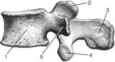
Fig. 2.5. Lumbar vertebra (lateral aspect):
Читать дальшеИнтервал:
Закладка:
Похожие книги на «Anatomy of bone system. The manual for medical students / Анатомия костной системы. Учебное пособие для медицинских вузов»
Представляем Вашему вниманию похожие книги на «Anatomy of bone system. The manual for medical students / Анатомия костной системы. Учебное пособие для медицинских вузов» списком для выбора. Мы отобрали схожую по названию и смыслу литературу в надежде предоставить читателям больше вариантов отыскать новые, интересные, ещё непрочитанные произведения.
![Евгений Аринин - Религиоведение [учебное пособие для студентов ВУЗов]](/books/106186/evgenij-arinin-religiovedenie-uchebnoe-posobie-dlya-thumb.webp)


Обсуждение, отзывы о книге «Anatomy of bone system. The manual for medical students / Анатомия костной системы. Учебное пособие для медицинских вузов» и просто собственные мнения читателей. Оставьте ваши комментарии, напишите, что Вы думаете о произведении, его смысле или главных героях. Укажите что конкретно понравилось, а что нет, и почему Вы так считаете.