Иван Гайворонский - Anatomy of bone system. The manual for medical students / Анатомия костной системы. Учебное пособие для медицинских вузов
Здесь есть возможность читать онлайн «Иван Гайворонский - Anatomy of bone system. The manual for medical students / Анатомия костной системы. Учебное пособие для медицинских вузов» — ознакомительный отрывок электронной книги совершенно бесплатно, а после прочтения отрывка купить полную версию. В некоторых случаях можно слушать аудио, скачать через торрент в формате fb2 и присутствует краткое содержание. ISBN: , Жанр: Биология, Детская образовательная литература, foreign_language, на английском языке. Описание произведения, (предисловие) а так же отзывы посетителей доступны на портале библиотеки ЛибКат.
- Название:Anatomy of bone system. The manual for medical students / Анатомия костной системы. Учебное пособие для медицинских вузов
- Автор:
- Жанр:
- Год:неизвестен
- ISBN:978-5-299-00639-1
- Рейтинг книги:4 / 5. Голосов: 1
-
Избранное:Добавить в избранное
- Отзывы:
-
Ваша оценка:
Anatomy of bone system. The manual for medical students / Анатомия костной системы. Учебное пособие для медицинских вузов: краткое содержание, описание и аннотация
Предлагаем к чтению аннотацию, описание, краткое содержание или предисловие (зависит от того, что написал сам автор книги «Anatomy of bone system. The manual for medical students / Анатомия костной системы. Учебное пособие для медицинских вузов»). Если вы не нашли необходимую информацию о книге — напишите в комментариях, мы постараемся отыскать её.
Структура пособия соответствует современным стандартам медицинского образования в России и важнейшим Европейским стандартам. Английская и латинская терминология приведены в соответствии с Международной анатомической номенклатурой.
Anatomy of bone system. The manual for medical students / Анатомия костной системы. Учебное пособие для медицинских вузов — читать онлайн ознакомительный отрывок
Ниже представлен текст книги, разбитый по страницам. Система сохранения места последней прочитанной страницы, позволяет с удобством читать онлайн бесплатно книгу «Anatomy of bone system. The manual for medical students / Анатомия костной системы. Учебное пособие для медицинских вузов», без необходимости каждый раз заново искать на чём Вы остановились. Поставьте закладку, и сможете в любой момент перейти на страницу, на которой закончили чтение.
Интервал:
Закладка:
1. Osteomalacia – disorder of calcification of newly formed osseous tissue which is caused by deficiency in calcium or vitamin D.
2. Osteoporosis – disorder of formation of the bony matrix during the skeleton formation (insufficient compensation of resorbed bone tissue). It occurs in elderly and old age and is caused by exessive resorption of osseous tissue.
3. Ectopic osteogenesis – ossification of soft tissues in abnormal places (in walls of arteries, kidneys etc.)
TEST QUESTIONS
1. What function does the skeleton carry out?
2. Name the types and functions of the bone marrow.
3. List the principles of the bone classification.
4. Give the characteristic of the primary and secondary bones.
5. What organic and inorganic substances are included into the composition of the bone (in what ratio)?
6. What connective tissue structure covers the bone from outside? What is its function?
7. What is the structural unit of osseous tissue? Name the types of osteocytes.
CLINICOANATOMICAL PROBLEM
During the surgical operation in a 10-year-old patient the metaepiphysial cartilage which separates the head of humerus from the body of humerus, was radically removed. What is the prognosis?
2 . SKELETON OF TRUNK
The skeleton of trunk consists of the vertebral column, or backbone, columna vertebralis , and the thorax, cavea thoracis (thorax) .
The vertebral column in an adult person consists of 24 individual vertebrae, sacrum and coccyx. There are the cervical vertebrae (7), thoracic (12) and lumbar ones (5) distinguished in the vertebral column. The sacrum consists of five fused sacral vertebrae. The coccyx consists of 3–5 fused coccygeal vertebrae. The thorax consists of 12 pairs of ribs with corresponding thoracic vertebrae and the sternum.
2.1. General Vertebral Features
The vertebra, vertebra , is comprised of the vertebral body, corpus vertebrae , anteriorly, the vertebral arch, arcus vertebrae , posteriorly and vertebral processes , processus vertebrae . The vertebral body is anterior and supporting part of the vertebra. The arch is located behind the vertebral body. The vertebral arch is attached to the body with the help of two pedicles of vertebral arch, pediculi arcus vertebrae , thus bounding the vertebral foramen, foramen vertebrale (fig. 2.1).
The foramina of all vertebrae form the vertebral canal, canalis vertebralis , enclosing the spinal cord. Openings for blood vessels named nutrient foramina, foramina nutricia , are visible on the surface of the vertebral body.
Seven processes project from the vertebral arch. An unpaired spinous process, processus spinosus , projects dorsally along the median line. The paired transverse processes, processus transversus , project on the right and on the left in the frontal plane. The paired superior and inferior articular processes, processus articularis superior et processus articularis inferior , project up and down from the arch. The bases of the articular processes bound the superior and inferior vertebral notches, incisura vertebralis superior et incisura vertebralis inferior . The inferior notches are deeper than the superior ones. When the vertebrae are articulated with each other, the inferior and superior notches form an intervertebral foramen, foramen intervertebrale , on the right and on the left. The intervertebral foramina transmit spinal nerves and blood vessels.
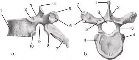
Fig. 2.1. Thoracic vertebra:
a – lateral aspect: 1 – vertebral body ( corpus vertebrae ); 2 – superior costal demi-facet ( fovea costalis superior ); 3 – superior vertebral notch ( incisura vertebralis superior ); 4 – superior articular process ( processus articularis superior ); 5 – transverse process ( processus transversus ); 6 – transverse costal facet ( fovea costalis processus transversi ); 7 – spinous process ( processus spinosus ); 8 – inferior articular process ( processus articularis inferior ); 9 – inferior vertebral notch ( incisura vertebralis inferior ); 10 – inferior costal demi-facet ( fovea costalis inferior )
b – superior aspect: 1 – spinous process ( processus spinosus ); 2 – vertebral arch ( arcus vertebrae ); 3 – pedicles of vertebral arch ( pediculi arcus vertebrae ); 4 – superior costal demi-facet ( fovea costalis superior ); 5 – superior articular process ( processus articularis superior ); 6 – transverse costal facet ( fovea costalis processus transversi ); 7 – transverse process ( processus transversus )
2.2. Cervical Vertebrae
The cervical vertebrae, vertebrae cervicales (C I– C VII), form the upper (cervical) part of the vertebral column. Two upper cervical vertebrae of seven significantly differ from other ones, therefore, they are termed atypical vertebrae. We will study them later. The other five vertebrae are structured according to the general principle: their bodies are relatively small, have an ellipsoid form, the vertebral foramen is large and triangular.
The distinctive feature of all cervical vertebrae is the presence of transverse foramina, foramen transversarium , in the transverse processes. They are formed as a result of the fusion of the transverse processes and the rudiments of the cervical ribs. The vertebral artery and vein pass through these foramina.
The groove for spinal nerve, sulcus nervi spinalis , passes along the superior surface of the transverse prosesses of the III–VII cervical vertebrae. These processes end with two tubercles – anterior and posterior, tuberculum anterius et tuberculum posterius . The anterior tubercle of the VI vertebra is more developed than the anterior tubercles of other cervical vertebrae. It is termed carotid tubercle, tuberculum caroticum , because the carotid artery can be compressed to it during hemorrhage.
The spinous processes are short and directed downwards, and their ends are bifurcated. The spinous process of the VII cervical vertebra is the longest, and its end is thickened. This vertebra is called prominent, vertebra prominens , because the apex of its spinous process is clearly palpated in a living person.
The articular processes of the cervical vertebrae are short and located obliquely between the frontal and horizontal planes. Meanwhile, the superior articular processes are directed backwards and slightly upwards, the inferior articular processes are directed forwards and slightly downwards (fig. 2.2).
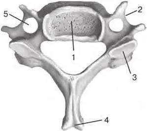
Fig. 2.2. Typical cervical vertebra (superior aspect):
1 – vertebral body ( corpus vertebrae ); 2 – transverse process ( processus transversus ); 3 – superior articular process ( processus articularis superior ); 4 – spinous process ( processus spinosus ); 5 – transverse foramen ( foramen transversarium )
The shape of the first two cervical vertebrae is influenced by their close location to the skull. They are involved in head turning. Therefore, they are termed «rotational vertebrae».
The I cervical vertebra is called the atlas, atlas (C I). It differs from the general structure of individual vertebrae: it has no body no notches and no spinous or articular processes (fig. 2.3).
Читать дальшеИнтервал:
Закладка:
Похожие книги на «Anatomy of bone system. The manual for medical students / Анатомия костной системы. Учебное пособие для медицинских вузов»
Представляем Вашему вниманию похожие книги на «Anatomy of bone system. The manual for medical students / Анатомия костной системы. Учебное пособие для медицинских вузов» списком для выбора. Мы отобрали схожую по названию и смыслу литературу в надежде предоставить читателям больше вариантов отыскать новые, интересные, ещё непрочитанные произведения.
![Евгений Аринин - Религиоведение [учебное пособие для студентов ВУЗов]](/books/106186/evgenij-arinin-religiovedenie-uchebnoe-posobie-dlya-thumb.webp)


Обсуждение, отзывы о книге «Anatomy of bone system. The manual for medical students / Анатомия костной системы. Учебное пособие для медицинских вузов» и просто собственные мнения читателей. Оставьте ваши комментарии, напишите, что Вы думаете о произведении, его смысле или главных героях. Укажите что конкретно понравилось, а что нет, и почему Вы так считаете.