Иван Гайворонский - Anatomy of bone system. The manual for medical students / Анатомия костной системы. Учебное пособие для медицинских вузов
Здесь есть возможность читать онлайн «Иван Гайворонский - Anatomy of bone system. The manual for medical students / Анатомия костной системы. Учебное пособие для медицинских вузов» — ознакомительный отрывок электронной книги совершенно бесплатно, а после прочтения отрывка купить полную версию. В некоторых случаях можно слушать аудио, скачать через торрент в формате fb2 и присутствует краткое содержание. ISBN: , Жанр: Биология, Детская образовательная литература, foreign_language, на английском языке. Описание произведения, (предисловие) а так же отзывы посетителей доступны на портале библиотеки ЛибКат.
- Название:Anatomy of bone system. The manual for medical students / Анатомия костной системы. Учебное пособие для медицинских вузов
- Автор:
- Жанр:
- Год:неизвестен
- ISBN:978-5-299-00639-1
- Рейтинг книги:4 / 5. Голосов: 1
-
Избранное:Добавить в избранное
- Отзывы:
-
Ваша оценка:
Anatomy of bone system. The manual for medical students / Анатомия костной системы. Учебное пособие для медицинских вузов: краткое содержание, описание и аннотация
Предлагаем к чтению аннотацию, описание, краткое содержание или предисловие (зависит от того, что написал сам автор книги «Anatomy of bone system. The manual for medical students / Анатомия костной системы. Учебное пособие для медицинских вузов»). Если вы не нашли необходимую информацию о книге — напишите в комментариях, мы постараемся отыскать её.
Структура пособия соответствует современным стандартам медицинского образования в России и важнейшим Европейским стандартам. Английская и латинская терминология приведены в соответствии с Международной анатомической номенклатурой.
Anatomy of bone system. The manual for medical students / Анатомия костной системы. Учебное пособие для медицинских вузов — читать онлайн ознакомительный отрывок
Ниже представлен текст книги, разбитый по страницам. Система сохранения места последней прочитанной страницы, позволяет с удобством читать онлайн бесплатно книгу «Anatomy of bone system. The manual for medical students / Анатомия костной системы. Учебное пособие для медицинских вузов», без необходимости каждый раз заново искать на чём Вы остановились. Поставьте закладку, и сможете в любой момент перейти на страницу, на которой закончили чтение.
Интервал:
Закладка:
After birth, reticulofibrous tissue is replaced with lamellar tissue formed of osteal lamellae 4,5 – 11 mkm thick. There are osteal cells (osteocytes) in the smallest cavities (lacunae) between osteal lamellae. Collagen fibers in the bone lamellae are strictly arranged parallelly to their surfaces. They lose connection with the connective tissue surrounding the bone. They are connected with the periosteum only due to perforating (Sharpey`s) fibers running from the periosteum to the superficial layers of the bone. The lamellar bone is much more solid than the reticulofibrous bone. Substitution of one osseous tissue with another is caused by the influence of functional loads on the skeleton. On the section of a macerated bone (bone deprived of soft tissues), it is possible to see two types of the osseous tissue: compact and spongy. The compact bone, substantia compacta , is a solid bony mass located on the exterior of the bone. The osteal lamellae of the compact bone are very close to each other. The compact tissue coats the epiphyses of tubular and flat bones as a thin sheath. The diaphyses of tubular bones entirely consist of compact bone.
Spongy bone, substantia spongiosa , is formed by loosely located osteal lamellae. In the spaces between them there is red bone marrow. Spongy tissue forms epiphyses of tubular bones, bodies of vertebrae, ribs, sternum, pelvic bones and some hand and foot bones. Only the superficial cortical layer of such bones is comprised of the compact tissue. The spongy tissue of the skull bones has significantly smaller regularly shaped spaces in comparison with the trunk and limb bones. It has a specific name – diploё.
The structural and functional unit of the bone is osteon or Haversian system. It is possible to see osteons on thin sections or on histological preparations. The osteon is formed of concentrically arranged osteal (Haversian) lamellae which surround the Haversian canal in the form of cylinders of various sizes nested into each other. The Haversian canal contains blood vessels and nerves. The majority of osteons are oriented parallelly to the axis of the bone joining with each other in many points. The number of osteons is individual for each bone. For example, in the femoral bone this number is 1,8 per 1 mm 2. Meanwhile, the share of the Haversian canal is 0,2 – 0,3 mm 2. Between the osteons there are insert or intermediate lamellae which run in all directions. The intermediate lamellae are the remnants of destroyed old osteons. Processes of new formation and destruction of osteons continuously occur in bones.
There is a layer of internal circumferential lamellae, lamina circumferentialis interna in tubular bones on the border with the medullary cavity. They are permeated with numerous canals widening to spaces. Several layers of general (or external) circumferential lamellae, lamina circumferentialis externa , surround the bone on the outside. The perforating canals (Volcmann`s canals) containing blood vessels with the same name pass through them.
There are three types of osteal lamellae in the diaphyses of tubular bones: Haversian, intermediate and general (external and internal). The lamellae lie close to each other, they are located parallelly to the axis of the bone and form quite a thick layer of only compact bone. It is 1,5 – 5 mm thick. Thus the diahysis of a long tubular bone is a hollow cylinder with walls formed of compact bone. The cavity of this cylinder is termed medullary canal. The latter is connected with spaces of the spongy tissue in the epiphyses of the bone. The Haversian lamellae form the basic mass of compact bone, thus making up osteons. The intermediate lamellae fill in the gaps between osteons. External and internal general (circumferential) lamellae form the most outer and the most inner layers of the compact bone, being located parallelly to the bone surface under the periosteum and endosteum respectively.
The epyphises of tubular bones consist of spongy tissue which is also formed of osteal lamellae. In structure, spongy bone may have large and small spaces. There are red bone marrow and vessels in these spaces. Compact bone covers epiphyses only on the outside with a comparatively thin layer. Flat and volumetric bones have a similar structure. Lamellae of spongy substance are strictly arranged in each bone. Their direction coincides with that of the maximum compression and stretching forces. The environment of each bone determines its structure. Trabeculae form an integral system in several adjacent bones, which characterizes the trabeculae’s architectonics. Such structure of bones preconditions their maximum solidity. In vertebrae, the stretching and compression forces are perpendicular to the superior and inferior surfaces of the vertebral bodies. This corresponds to the fact that the trabeculae have mainly vertical direction in spongy substance (fig. 1.2). In the proximal epiphysis of the femoral bone there are arch-shaped systems of trabeculae which transfer pressure from the surface of the bone head to the walls of the diaphysis. Besides, there are trabeculae transfering the traction force of muscles attached to the greater trochanter (fig. 1.3).
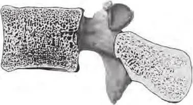
Fig. 1.2. Orientation of trabeculae in the vertebral body (saggital section)
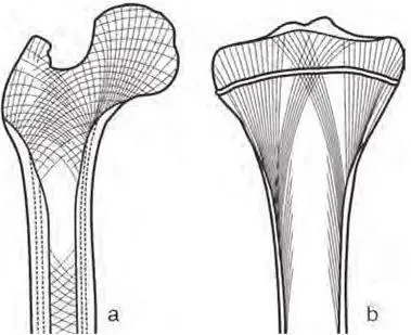
Fig. 1.3. Orientation of trabeculae in proximal epiphyses of tubular bones: a – in femur; b – in tibia
Trabeculae running in the radial direction are typical of the calcaneus. They distribute loads equally over the surface of the calcaneal tuberosity which serves as a foot support (fig. 1.4).
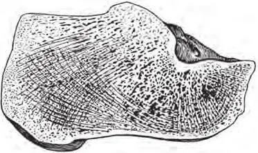
Fig. 1.4. Orientaton of trabeculae in calcaneus
Compact bone is formed in places of the highest concentration of force trajectories. It is clearly visible on the section of the femoral, tibial and calcaneal bones where the compact tissue is thickened in the areas of crossing between force lines and the bone surface. Thus we can say that compact bone is the result of compression of spongy bone, and vice versa, it is possible to consider spongy bone as sparse compact bone. It should be noted that if static and dynamic conditions are changed (increase or decrease in functional loads), the spongy bone architectonics changes too, a part of trabeculae disappear, or new systems of osteal trabeculae develop. The spongy bone structure changes in a special visible manner after fractures.
1.4. External Structure of Bones
While describing the external structure of bones, we should pay attention to the surfaces, facies , of the bones, which may be flat, concave or convex, smooth or rough. Articular surfaces facies articularis , involved in formation of joints, are the most smoothly polished ones. In some bones the end is rounded, forming a head – caput ; at the same time, the end of other bones has concavity, called articular fossa, or fossa articularis . The head may be separated from the bone body with a constricted part – neck, collum . If the articular end is extensive but slightly curved surface, it is termed condyle, condilus . The processes located near the condyle are named epicondyles, epicondyli, they serve for attachment of tendons and ligaments (they may also be called apophyses).
The following surfaces are distinguished in bones (depending on theirlocation in the human body): internal or external, medial or lateral etc. The surfaces are separated by borders, margo . The borders, in turn, are known as superior or inferior, medial or lateral etc. They may be smooth or serrated, blunt or sharp, sometimes they have notches, incisurae , of different sizes.
Читать дальшеИнтервал:
Закладка:
Похожие книги на «Anatomy of bone system. The manual for medical students / Анатомия костной системы. Учебное пособие для медицинских вузов»
Представляем Вашему вниманию похожие книги на «Anatomy of bone system. The manual for medical students / Анатомия костной системы. Учебное пособие для медицинских вузов» списком для выбора. Мы отобрали схожую по названию и смыслу литературу в надежде предоставить читателям больше вариантов отыскать новые, интересные, ещё непрочитанные произведения.
![Евгений Аринин - Религиоведение [учебное пособие для студентов ВУЗов]](/books/106186/evgenij-arinin-religiovedenie-uchebnoe-posobie-dlya-thumb.webp)


Обсуждение, отзывы о книге «Anatomy of bone system. The manual for medical students / Анатомия костной системы. Учебное пособие для медицинских вузов» и просто собственные мнения читателей. Оставьте ваши комментарии, напишите, что Вы думаете о произведении, его смысле или главных героях. Укажите что конкретно понравилось, а что нет, и почему Вы так считаете.