O. Belousova - Pediatric stroke. Revascularization and reconstructive surgery in children with cerebrovascular disease
Здесь есть возможность читать онлайн «O. Belousova - Pediatric stroke. Revascularization and reconstructive surgery in children with cerebrovascular disease» — ознакомительный отрывок электронной книги совершенно бесплатно, а после прочтения отрывка купить полную версию. В некоторых случаях можно слушать аудио, скачать через торрент в формате fb2 и присутствует краткое содержание. ISBN: , Жанр: russian_contemporary, Медицина, на английском языке. Описание произведения, (предисловие) а так же отзывы посетителей доступны на портале библиотеки ЛибКат.
- Название:Pediatric stroke. Revascularization and reconstructive surgery in children with cerebrovascular disease
- Автор:
- Жанр:
- Год:неизвестен
- ISBN:9785449342652
- Рейтинг книги:5 / 5. Голосов: 1
-
Избранное:Добавить в избранное
- Отзывы:
-
Ваша оценка:
Pediatric stroke. Revascularization and reconstructive surgery in children with cerebrovascular disease: краткое содержание, описание и аннотация
Предлагаем к чтению аннотацию, описание, краткое содержание или предисловие (зависит от того, что написал сам автор книги «Pediatric stroke. Revascularization and reconstructive surgery in children with cerebrovascular disease»). Если вы не нашли необходимую информацию о книге — напишите в комментариях, мы постараемся отыскать её.
Pediatric stroke. Revascularization and reconstructive surgery in children with cerebrovascular disease — читать онлайн ознакомительный отрывок
Ниже представлен текст книги, разбитый по страницам. Система сохранения места последней прочитанной страницы, позволяет с удобством читать онлайн бесплатно книгу «Pediatric stroke. Revascularization and reconstructive surgery in children with cerebrovascular disease», без необходимости каждый раз заново искать на чём Вы остановились. Поставьте закладку, и сможете в любой момент перейти на страницу, на которой закончили чтение.
Интервал:
Закладка:
Radiation-induced vasculopathies with subsequent vascular stenosis or occlusion more often occur in patients with brain tumors. A recent study showed that after radiation therapy courses the radiographic signs of strokes were noted approximately in 6% of children with tumors of the central nervous system [92].
One of the causes of brain blood supply disturbance in children is a cerebrovascular pathology . Presently, many authors come to the conclusion of an imperative need in a more detailed examination of children for presence of cerebrovascular diseases. In the situations, which require a compensation by means of the blood flow redistribution (e.g., during a prolonged head tilt), the decisive role may be played by specific anatomic features of major arteries of the head and the neck as well as of the basilar vessels, which can often be found even in healthy people (non-closed Willis’ circle, hypoplasias of individual artery segments or of whole arteries, stenosis / occlusion of arteries, pathological deformation of ICA) [7; 12; 30; 108].
The prevalence of ischemic disturbances, which, as a rule, are preceded by a chronic cerebrovascular insufficiency, in the structure of the young age stroke requires a comprehensive study of age peculiarities in the cerebrovascular system functioning. In principle, the brain blood supply physiology in children does not differ from that of adults. For instance, a brain weight is only 2% of the total weight of man, but it receives about 20% of the blood volume during the cardiac output and consumes 20% of oxygen. An adequate cerebral blood flow sufficient for sustaining a normal life activity of the brain is, on the average, 45—60 mL/100 g of brain tissue per minute. The proper brain activity depends on regular and adequate blood supply due to its high metabolic activity and lack of any significant energy reserves. The state of the cerebral blood flow insufficiency known as a cerebral ischemia may be either acute, or chronic and may have either local or widespread nature. When the blood flow decreases to 20—35 mL/100 g/min., the electrical activity of the brain drops, but the brain tissue changes are reversible. This state is often defined as a «penumbra» – an ischemic penumbra. When the blood flow decreases to 10—15 mL/100 g/min., the changes developing in the brain tissues become irreversible. This process leads to the death of cells (brain infarction). Timely restored adequate cerebral blood flow is enormously important in treatment of a specific group of patients with a chronic cerebral ischemia caused by a pathology of major cerebral vessels [92]. The study of cerebrovascular disturbances may turn to be a connecting link between a cerebrovascular pathology in children and the subsequent development of strokes in adults.
The anatomy of brachiocephalic arteries varies in individuals. Both in children and adults, the Willis’ circle is formed correctly only in 18—20%. Many authors think that the anatomy of cerebral vessels does not depend on gender, age or race [89; 150]. In the opinions of other authors, in children the specific anatomic and physiological features of BCA and intracranial arteries take shape, mainly, in the younger age group. For instance, the collateral vessels between ECA and ICA via a. ophtalmica are formed only in 17—22% of children [173]. The morphometric parameters of ICA grow in school-aged children from 7 to 18 years old. The morphometric parameters of common carotid arteries grow leapwise. In girls, the maximum growth occurs earlier than in boys [6].
More than a century ago in literature there were already mentions made of specific anatomical features of ICA structure – tortuosity, kink and looping. Many anatomists described bent, deformed ICA. W. Coulson (1852) was, probably, the first, who mentioned the ICA looping describing it as «a pulsating tumor on the neck». The connection between the pathologic deformations and the high risk of the CVD development was first noted by M. Riser et al. in 1951 [218]. F. McDowell et al. (1959) reported 20 cases of pathological tortuosity of ICA and related blood flow disturbances, when studying 68 patients with cerebrovascular pathology among adults over 30 years old without any proofs of occlusion found, which was supported by angiography [92]. H. Metz et al. (1961) held a retrospective survey of 1000 angiograms and found 161 cases of kinks and looping on ICA, including children below 10 years old [182]. In 1965 after having described 2,453 angiograms J. Weibel and W. Fields revealed a high occurrence of this specific feature of anatomical structure in the population – up to 10—15% cases [274]. N. Sarkari et al. (1970) first described 8 children with the pathological tortuosity of ICA combined with a chronic cerebral ischemia, of which 7 children were below 10 years old [228].
The development of contemporary diagnostic methods led to more frequent detection of ICA deformations in a population. According to US test data, the detection rate of ICA tortuosity reaches 25—30% in adults and 43% in children (R. Hobson, 2004). According to data of other authors (E. Ballota et al. (2005), G. La Barbera et al. (2006), W. Perdue et al. (1975), C. Togay-Isikay et al. (2005)), pathological deformations occur in 10—40% of cases depending on the population surveyed.
In the opinion of W. Fisher (1982), the surgical treatment of cerebrovascular insufficiency in children is required in cases, when the ICA changes may lead to a progressing neurologic deficiency. The author also states that cerebrovascular insufficiency in children presents a serious problem, as an inadequate perfusion of brain tissue leads to irreversible changes with the subsequent formation of a neurologic deficiency. In his opinion, the most frequent causes leading to cerebrovascular insufficiency are the following ICA changes: 1) congenital pathological tortuosity (kinking), 2) post-traumatic changes with thrombosis, aneurysm and embolism, and 3) thromboses [97].
In children, CVDs may be also caused by: the moya-moya disease, a fibro-muscular dysplasia, an artery dissection [8; 20]. All the above-mentioned diseases lead either to stenosis or luminal occlusion [213].
Fibro-muscular dysplasia is one of the causes of a pathological deformation of the ICA and a pediatric stroke. The pathomorphology of the fibro-muscular dysplasia is characterized by the emergence of hyperplasia sites on the connective tissue, an irregular atrophy of muscular fibers and degenerative changes of vascular walls. All this leads to formation of stenosis sites interspersed with post-stenotic aneurysms. This variant of impairment of brachiocephalic vessels may be isolated and generalized, more frequently unilateral, although a bilateral impairment is not ruled out [211].
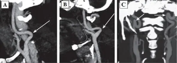
Fig. 2:Types of pathologic deformations according to SCT AG data: A – Type 1 septal stenosis (angle over 60°) (arrow indicated); B – Type 2 septal stenosis (angle from 30 to 60°) (arrow indicated); C – Type 3 septal stenosis (angle less than 30°) (arrow indicated).
The permanent effect of the force vector of arterial blood pressure on dysplastic vascular wall was discussed earlier as the most intensive initiating agent forming a pathologic tortuosity of the ICA [145]. In 1961 H. Metz offered his own classification of ICA deformations. The author supposed that the intensity of the process depended on dysplastic changes of the vascular wall and vessel configuration and divided septal stenoses of the ICA into three types:
Type 1: artery kink at an angle over 60°(Fig. 2A);
Type 2: artery kink at an angle from 30 to 60°(Fig. 2B);
Читать дальшеИнтервал:
Закладка:
Похожие книги на «Pediatric stroke. Revascularization and reconstructive surgery in children with cerebrovascular disease»
Представляем Вашему вниманию похожие книги на «Pediatric stroke. Revascularization and reconstructive surgery in children with cerebrovascular disease» списком для выбора. Мы отобрали схожую по названию и смыслу литературу в надежде предоставить читателям больше вариантов отыскать новые, интересные, ещё непрочитанные произведения.


Обсуждение, отзывы о книге «Pediatric stroke. Revascularization and reconstructive surgery in children with cerebrovascular disease» и просто собственные мнения читателей. Оставьте ваши комментарии, напишите, что Вы думаете о произведении, его смысле или главных героях. Укажите что конкретно понравилось, а что нет, и почему Вы так считаете.