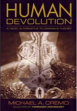
Michael Cremo - Human Devolution - A Vedic Alternative To Darwin's Theory
Здесь есть возможность читать онлайн «Michael Cremo - Human Devolution - A Vedic Alternative To Darwin's Theory» весь текст электронной книги совершенно бесплатно (целиком полную версию без сокращений). В некоторых случаях можно слушать аудио, скачать через торрент в формате fb2 и присутствует краткое содержание. Год выпуска: 2003, ISBN: 2003, Издательство: Torchlight Publishing, Жанр: Старинная литература, на английском языке. Описание произведения, (предисловие) а так же отзывы посетителей доступны на портале библиотеки ЛибКат.
- Название:Human Devolution: A Vedic Alternative To Darwin's Theory
- Автор:
- Издательство:Torchlight Publishing
- Жанр:
- Год:2003
- ISBN:9780892133345
- Рейтинг книги:4 / 5. Голосов: 1
-
Избранное:Добавить в избранное
- Отзывы:
-
Ваша оценка:
- 80
- 1
- 2
- 3
- 4
- 5
Human Devolution: A Vedic Alternative To Darwin's Theory: краткое содержание, описание и аннотация
Предлагаем к чтению аннотацию, описание, краткое содержание или предисловие (зависит от того, что написал сам автор книги «Human Devolution: A Vedic Alternative To Darwin's Theory»). Если вы не нашли необходимую информацию о книге — напишите в комментариях, мы постараемся отыскать её.
Human Devolution: A Vedic Alternative To Darwin's Theory — читать онлайн бесплатно полную книгу (весь текст) целиком
Ниже представлен текст книги, разбитый по страницам. Система сохранения места последней прочитанной страницы, позволяет с удобством читать онлайн бесплатно книгу «Human Devolution: A Vedic Alternative To Darwin's Theory», без необходимости каждый раз заново искать на чём Вы остановились. Поставьте закладку, и сможете в любой момент перейти на страницу, на которой закончили чтение.
Интервал:
Закладка:
Darwin’s vague account of a light-sensitive spot gradually developing into the complex, cameralike human eye may have a certain superficial plausibility, but it does not constitute a scientific explanation of the eye’s origin. It is simply an invitation to imagine that evolution actually took place. If one wishes to turn imagination into science, one must take into account the structure of the eye on the biomolecular level.
Devlin (1992, pp. 938–954) gives a fairly detailed biochemical description of the human vision process. Biochemist Michael Behe (1996, pp. 18–21) summarizes Devlin’s explanation like this: “When light first strikes the retina a photon interacts with a molecule called 11- cis -retinal, which rearranges within picoseconds to trans -retinal. . . . The change in the shape of the retinal molecule forces a change in the shape of the protein, rhodopsin, to which the retinal is tightly bound. . . . Now called metarhodopsin II, the protein sticks to another protein, called transducin. Before bumping into metarhodopsin II, transducin had tightly bound a small molecule called GDP. But when transducin interacts with metarhodopsin II, the GDP falls off, and a molecule called GTP binds to transducin. . . . GTP-transducin-metarhodopsin II now binds to a protein called phosphodiesterase, located in the inner membrane of the cell. When attached to metarhodopsin II and its entourage, the phosphodiesterase acquires the chemical ability to ‘cut’ a molecule called cGMP . . . Initially there are a lot of cGMP molecules in the cell, but the phosphodiesterase lowers its concentration, just as a pulled plug lowers the water level in a bathtub. Another membrane protein that binds cGMP is called an ion channel. It acts as a gateway that regulates the number of sodium ions in the cell, while a separate protein actively pumps them out again. The dual action of the ion channel and pump keeps the level of sodium ions in the cell within a narrow range. When the amount of cGMP is reduced because of cleavage by the phosphodiesterase, the ion channel closes, causing the cellular concentration of positively charged sodium ions to be reduced. This causes an imbalance of charge across the cell membrane that, finally, causes a current to be tranmitted down the optic nerve to the brain. The result, when interpreted by the brain, is vision.”
Another equally complex set of reactions restores the original chemical elements that started the process, like 11- cis -retinal, cGMP, and sodium ions (Behe 1996, p. 21). And this is just part of the biochemistry underlying the process of vision. Behe (1996, p. 22) stated: “Ultimately . . . this is the level of explanation for which biological science must aim. In order to truly understand a function, one must understand in detail every relevant step in the process. The relevant steps in biological processes occur ultimately at the molecular level, so a satisfactory explanation of a biological phenomenon—such as sight, digestion, or immunity—must include its molecular explanation.” Evolutionists have not produced such an explanation.
The vesicular transport System
The lysosome is a compartment within the cell that disposes of damaged proteins. There are enzymes within the lysosome that dismantle the proteins. These enzymes are manufactured in ribosomes, compartments found inside another cellular compartment called the endoplasmic reticulum. As the enzymes are being manufactured in the ribosomes, they are tagged with special amino acid sequences that allow them to pass through the walls of the ribosomes into the endoplasmic reticulum. From there, they are tagged with other amino acid sequences that allow them to pass out of the endoplasmic reticulum. The enzymes make their way to the lysosome, where they bind to the surface of the lysosome. Then yet another set of signal tags allow them to enter the lysosome, where they can do their work (Behe 1998, pp. 181–182; Alberts et al. 1994, pp. 551–650). This transportation network is called the vesicular transport system.
72 Human Devolution: a vedic alternative to Darwin’s theory
In I-cell disease, a flaw in signal tagging disrupts the vesicular transport system. Instead of carrying the protein-degrading enzymes from the ribosomes to the lysosomes, the system carries them to the cell wall, where they are dumped outside of the cell. Meanwhile, damaged proteins flow into the lysosomes, where they are not degraded. Without the proteindegrading enzymes, the lysosomes fill up like overflowing garbage cans. To deal with this, the cell manufactures new lysosomes, which also fill up with garbage proteins. Finally, when there are too many lysosomes filled with garbage proteins, the whole cell breaks down and the person with this disease dies. This shows what happens when one part of a complex system is missing—the whole system breaks down. All the parts of the vesicular transport system have to be in place for it to work effectively.
Behe (1996, pp. 115–116) says: “Vesicular transport is a mind-boggling process, no less complex than the completely automated delivery of vaccine from a storage area to a clinic a thousand miles away. Defects in vesicular transport can have the same deadly consequences as the failure to deliver a needed vaccine to a disease-racked city. An analysis shows that vesicular transport is irreducibly complex, and so its development staunchly resists gradualistic explanations, as Darwinian evolution would have it. A search of the professional biochemical literature shows that no one has ever proposed a detailed route by which such a system could have come to be. In the face of the enormous complexity of vesicular transport, Darwinian theory is mute.”
The Blood Clotting mechanism
The human blood clotting mechanism is another puzzle for evolutionists. Behe (1996, p. 78) says: “Blood clotting is a very complex, intricately woven system consisting of scores of interdependent protein parts. The absence of, or significant defects in, any one of a number of the components causes the system to fail: blood does not clot at the proper time or at the proper place.” The system is thus one of irreducible complexity, not easily explained in terms of Darwinian evolution.
The blood clotting mechanism centers around fibrinogen, a blood protein that forms the fibers that make up the clots. Normally, fibrinogen is dissolved in the blood plasma. When bleeding begins, a protein called thrombin cuts fibrinogen to make strings of a protein called fibrin. The fibrin filaments stick together, forming a network that catches blood cells, thus stopping the flow of blood from a wound (Behe 1996, p. 80). At first, the network is not very strong. It sometimes breaks, allowing the blood to flow out from the wound again. To prevent this, a protein called the fibrin stabilizing factor (FSF), creates cross links between fibrin filaments, strengthening the network (Behe 1996, p. 88).
Meanwhile, thrombin is cutting more fibrinogen into more fibrin, which forms more clots. At a certain point, the thrombin has to stop cutting fibrinogen or else so much fibrin would be produced that it would clot up the whole blood system and the person would die (Behe 1996, p.81).
There is a complex cascade of proteins and enzymes involved in turning the blood clotting system on and off at the proper times. Thrombin originally exists as an inactive form, prothrombin. In this form, it doesn’t cut fibrinogen into the fibrin filaments that make clots. So for the clotting process to start, prothrombin must be converted to thrombin. Otherwise, a person bleeds to death. And once the proper clotting is formed, thrombin has to be turned back into prothrombin. Otherwise, the clotting continues until all the blood stops flowing (Behe 1996, p. 82).
A protein called the Stuart factor is involved in the activation of prothrombin, turning it into thrombin, so that the clotting process can start. So what activates the inactive Stuart factor? There are two cascades of interactions, which begin with transformations at the wound site. Let’s consider just one of them. Behe (1996, p. 84) says: “When an animal is cut, a protein called Hageman factor is then cleaved by a protein called HMK to yield activated Hageman factor. Immediately the activated Hageman factor converts another protein, called prekallikrein, to its active form, kallikrein. Kallikrein helps HMK speed up the conversion of more Hageman factor to its active form. Activated Hageman factor and HMK then together transform another protein, called PTA, to its active form. Activated PTA in turn, together with the activated form of another protein called convertin, switch a protein called Christmas factor to its active form. Finally, activated Christmas factor, together with antihemophilic factor . . . changes Stuart to its active form.” The second cascade is equally complicated, and in some places merges with the first.
Читать дальшеИнтервал:
Закладка:
Похожие книги на «Human Devolution: A Vedic Alternative To Darwin's Theory»
Представляем Вашему вниманию похожие книги на «Human Devolution: A Vedic Alternative To Darwin's Theory» списком для выбора. Мы отобрали схожую по названию и смыслу литературу в надежде предоставить читателям больше вариантов отыскать новые, интересные, ещё непрочитанные произведения.
Обсуждение, отзывы о книге «Human Devolution: A Vedic Alternative To Darwin's Theory» и просто собственные мнения читателей. Оставьте ваши комментарии, напишите, что Вы думаете о произведении, его смысле или главных героях. Укажите что конкретно понравилось, а что нет, и почему Вы так считаете.


