Brenda A. Wilson - Bacterial Pathogenesis
Здесь есть возможность читать онлайн «Brenda A. Wilson - Bacterial Pathogenesis» — ознакомительный отрывок электронной книги совершенно бесплатно, а после прочтения отрывка купить полную версию. В некоторых случаях можно слушать аудио, скачать через торрент в формате fb2 и присутствует краткое содержание. Жанр: unrecognised, на английском языке. Описание произведения, (предисловие) а так же отзывы посетителей доступны на портале библиотеки ЛибКат.
- Название:Bacterial Pathogenesis
- Автор:
- Жанр:
- Год:неизвестен
- ISBN:нет данных
- Рейтинг книги:5 / 5. Голосов: 1
-
Избранное:Добавить в избранное
- Отзывы:
-
Ваша оценка:
Bacterial Pathogenesis: краткое содержание, описание и аннотация
Предлагаем к чтению аннотацию, описание, краткое содержание или предисловие (зависит от того, что написал сам автор книги «Bacterial Pathogenesis»). Если вы не нашли необходимую информацию о книге — напишите в комментариях, мы постараемся отыскать её.
Completely revised and updated, and for the first time in stunning full-color, Bacterial Pathogenesis: A Molecular Approach, Fourth Edition, builds on the core principles and foundations of its predecessors while expanding into new concepts, key findings, and cutting-edge research, including new developments in the areas of the microbiome and CRISPR as well as the growing challenges of antimicrobial resistance. All-new detailed illustrations help students clearly understand important concepts and mechanisms of the complex interplay between bacterial pathogens and their hosts. Study questions at the end of each chapter challenge students to delve more deeply into the topics covered, and hone their skills in reading, interpreting, and analyzing data, as well as devising their own experiments. A detailed glossary defines and expands on key terms highlighted throughout the book. Written for advanced undergraduate, graduate, and professional students in microbiology, bacteriology, and pathogenesis, this text is a must-have for anyone looking for a greater understanding of virulence mechanisms across the breadth of bacterial pathogens.
Bacterial Pathogenesis — читать онлайн ознакомительный отрывок
Ниже представлен текст книги, разбитый по страницам. Система сохранения места последней прочитанной страницы, позволяет с удобством читать онлайн бесплатно книгу «Bacterial Pathogenesis», без необходимости каждый раз заново искать на чём Вы остановились. Поставьте закладку, и сможете в любой момент перейти на страницу, на которой закончили чтение.
Интервал:
Закладка:
Skin consists of a lining of living cells composed of two layers, the epidermis (outer layer) and the dermis (inner layer) ( Figure 2-1). Cells of the dermis are continuously replaced as they die and become a tough, dry outer layer called the epidermis. The epithelia that line the respiratory, intestinal, and urogenital tracts are the mucosal epithelia and consist of tightly packed cells that are attached to each other by protein structures called tight adherens junctions and desmosomes ( Figure 2-2). The tight binding of epithelial cells to each other prevents bacteria from transiting an epithelial layer. To get through the epithelial layer, bacteria must either take advantage of wounds or be capable of invading epithelial cells, passing between or through them to get to the underlying tissue.
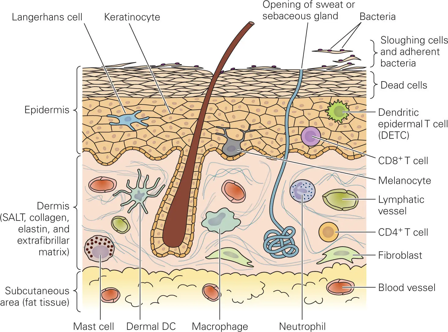
Figure 2-1. Structure of the skin. The thick epidermis (outer layer, top) consists of keratinized, stratified squamous cells, the top layers of which are dead and continuously shed. When these cells are sloughed, they carry adhering bacteria with them. The inner layer is the dermis (middle). Sebaceous (fat) glands, sweat glands, and hair follicles, which produce antibacterial substances, provide openings in the skin through which pathogenic bacteria occasionally enter. The skin also has an adaptive defense system, the skin-associated lymphoid tissue (SALT). Langerhans cells serve as phagocytic cells in this system.
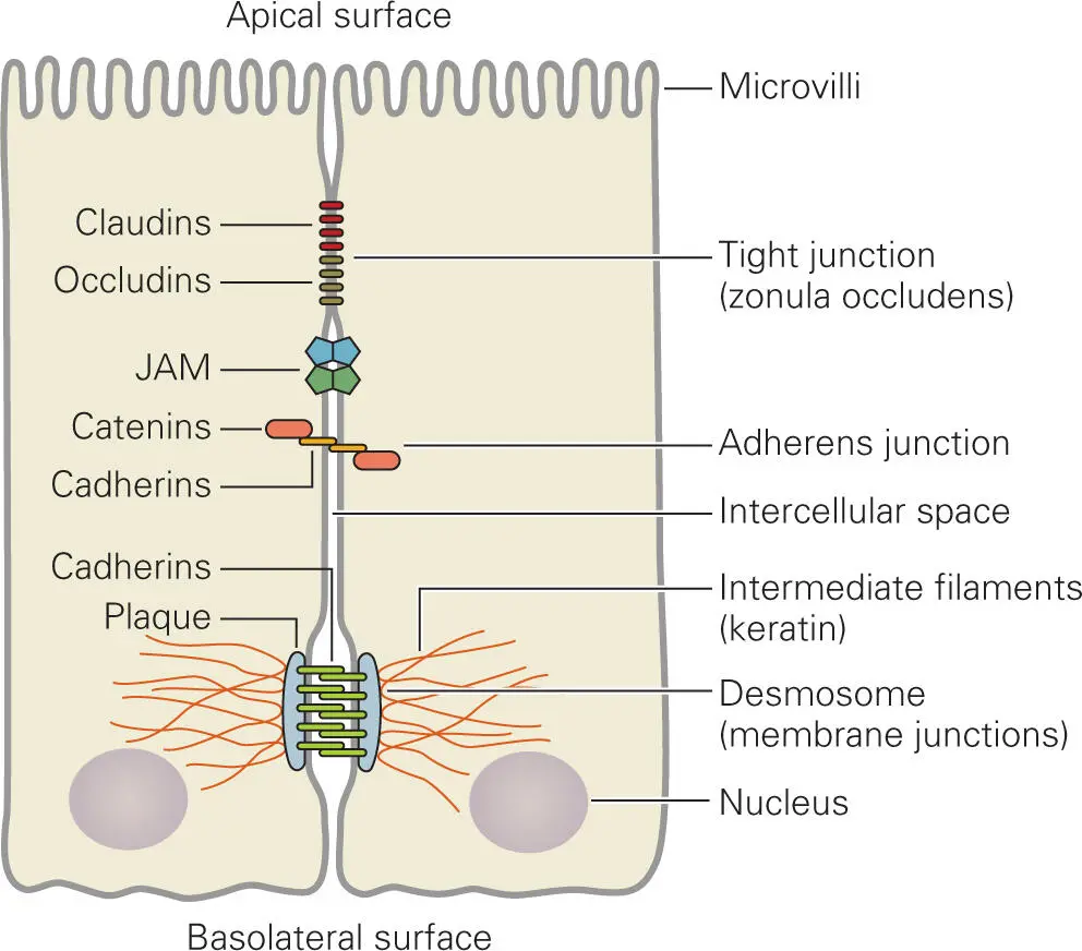
Figure 2-2. Intestinal epithelial cells showing tight, junctional, and adherens junctions, and desmosomes. JAM, junctional adhesion molecules.
In contrast, the cells that line the surfaces of the interior of the body (the endothelium), such as blood vessels or lymphatic vessels, are not tightly bound to each other in order to allow the cells of the immune defense system to move freely from blood to tissues. Unfortunately, this feature also allows bacteria to move into and out of blood and lymphatic vessels by moving between the endothelial cells. Thus, once bacteria gain entry into the body at one site, it is possible for them to readily gain access to other parts of the body. Because of this vulnerability, it is imperative that the epithelia of skin and mucosal surfaces function as barriers against foreign invaders.
The membrane surface of an epithelial cell that faces toward the interior tissues of the body and is attached to other cells or to connective tissues (basolateral surface) has a different protein composition from the membrane surface that faces outward (apical surface). Cells with this asymmetrical surface property are said to be polarized cells. A feature of epithelial cells is that they are attached to a thin sheet of connective tissue called the basement membrane (basal lamina). The basal lamina covers regions of loose connective tissue ( Figure 2-3), comprised of extracellular matrix (ECM) secreted by elongated fibroblast cells. The ECM composition varies with tissue type, but primarily contains a network of interlocking gels of polysaccharides called glycosaminoglycans (such as chondroitin sulfate, hyaluronan, keratan sulfate, and heparan sulfate) attached to fibrous collagens, the most abundant protein type in the ECM. Collagens bind to adhesion glycoproteins called fibronectins, which in turn bind collagens to transmembrane cell surface proteins called integrins that mediate cell-cell and cell-ECM interactions and cytoskeletal responses. Other ECM proteins called laminins bind to collagens and other ECM components to form fibrous networks that resist tensile forces in the basal lamina. There are numerous examples of pathogenic bacteria that attach to components of the ECM and manipulate or mimic ECM components during the course of infection.
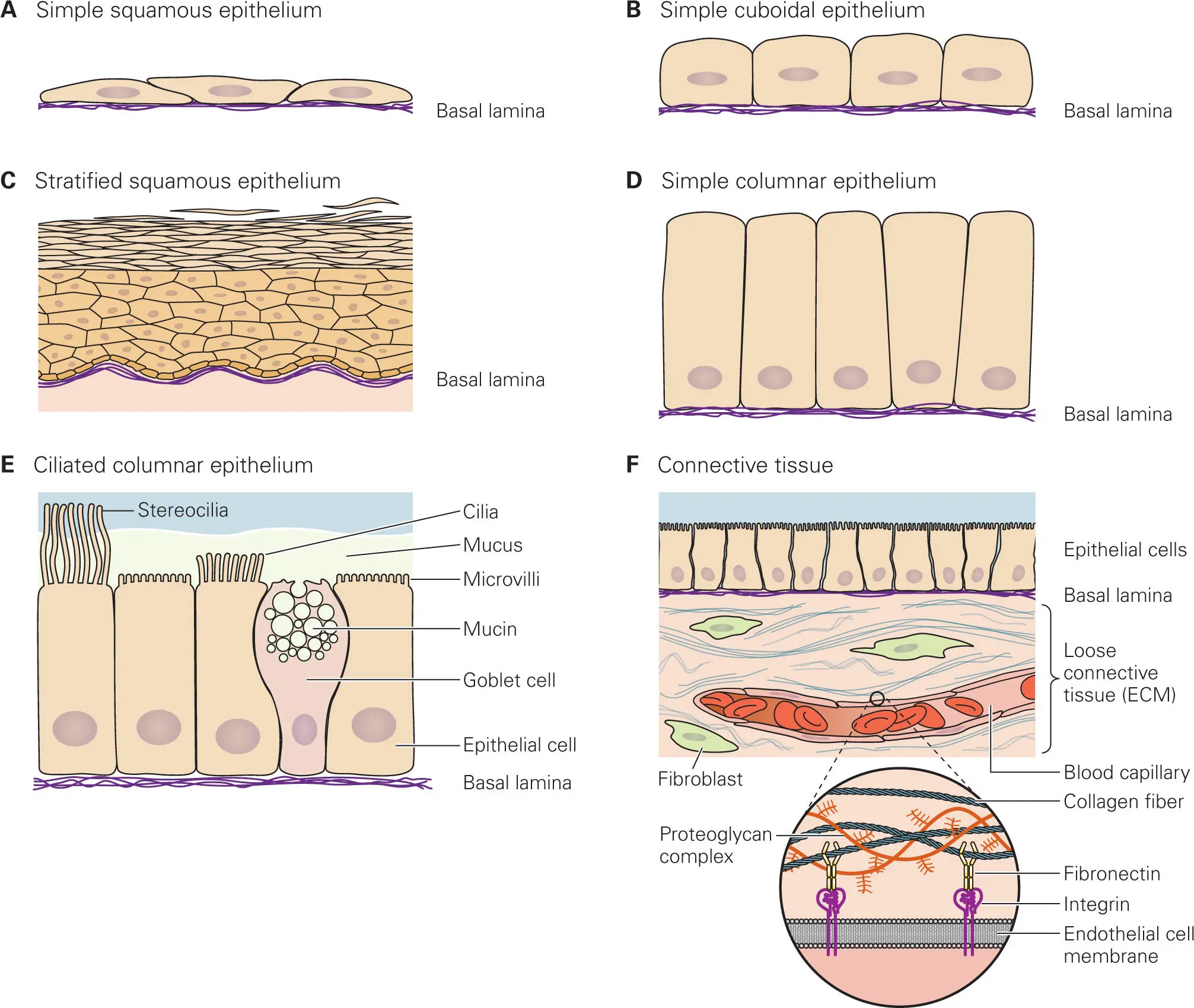
Figure 2-3. Different types of epithelial cells and their relationships to underlying tissue. Shown are (A) simple squamous epithelium; (B) simple cuboidal epithelium; (C) stratified squamous epithelium (upper layers of cells are dead, typical of skin); (D) simple columnar epithelium; (E) ciliated columnar epithelium showing goblet cells, which secrete mucus (mucin); and (F) typical structure of connective tissue under an epithelial cell layer. Panel F modified from Cooper GM, Hausman RE. 2007. The Cell—A Molecular Approach, 4th ed. ASM Press, Washington, DC.
Epithelial cells in different body sites vary in shape, size, and number of layers ( Figure 2-3), as well as in functional properties. Epithelial layers that cover surfaces where absorption or secretion is taking place (e.g., in the intestinal tract) usually consist of a single layer of epithelial cells (simple epithelium). Other surfaces, such as the female cervix or the skin, are composed of many layers of epithelial cells (stratified epithelium). Some have a flattened shape (squamous epithelium) and form the lining of cavities (such as the mouth, heart, and lungs) and the outer layers of the skin. Some are cube-shaped (cuboidal epithelium) and form the lining of kidney tubules and gland ducts and constitute the germinal epithelium that develops into egg and sperm cells. Others are tall and thin (columnar epithelium) and form the lining of the stomach and intestine.
Most of the surfaces that are exposed directly to the environment (e.g., the skin and mouth) are covered by stratified epithelia, whereas simple epithelia are found in internal areas, such as the intestinal tract or the lungs. Simple epithelia are more vulnerable to bacterial invasion than stratified epithelia because invading bacteria only have to pass through one layer of cells to gain access to the tissue underneath. We will use the terms mucosa epithelia, epithelial cells, mucosal layer, or mucosal cells to denote the simple epithelia of these internal areas.
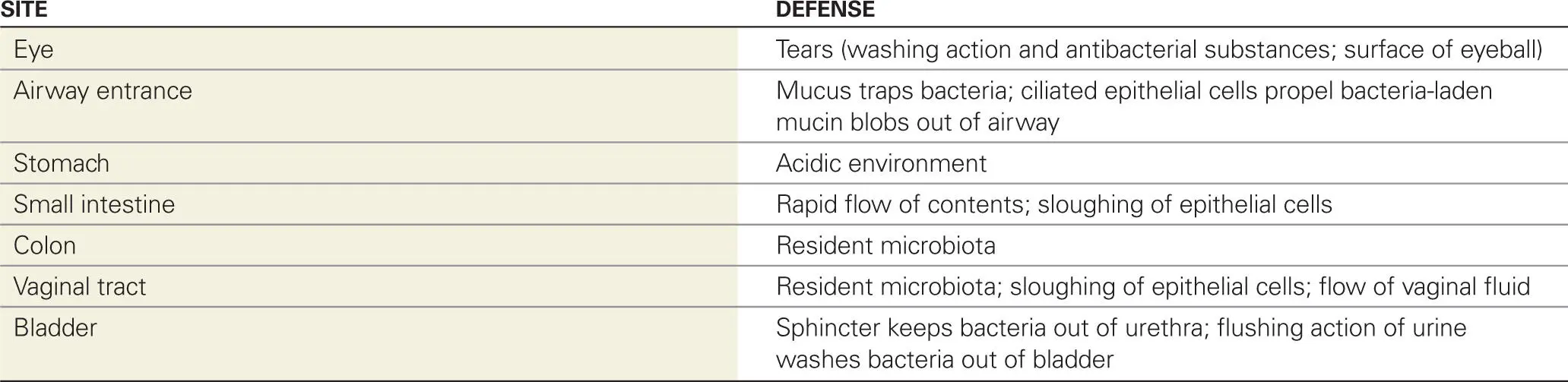
Epithelia are protected by an array of innate and adaptive defenses. Some of these defenses are listed in Table 2-1for the skin and Table 2-2for mucosal surfaces. Other defenses, listed in Table 2-3, are more specific to certain areas of the body, such as the eyes and the respiratory, gastrointestinal, and urogenital tracts. For example, tears contain an enzyme (lysozyme) that degrades bacterial cell walls. Tears also provide a washing action that removes particles and bacteria from the eyes. Entry to the respiratory tract is protected by mucus and by specialized ciliated cells that propel bacteria-laden blobs of mucus out of the lungs. The urinary tract epithelium is protected by a sphincter at the end of the urethra, the tube that leads up to the bladder. This barrier makes it difficult for bacteria to enter the tube that leads to the bladder. Also, the washing action of urine during urination flushes out any bacteria that may have gained access to the bladder and urethra.
Table 2-1. Defenses of the skin
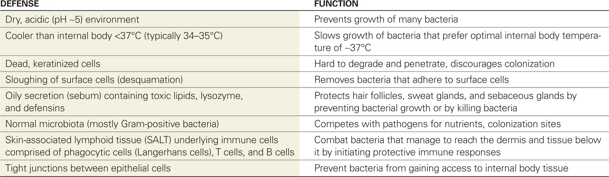
Table 2-2. Defenses of mucosal surfaces
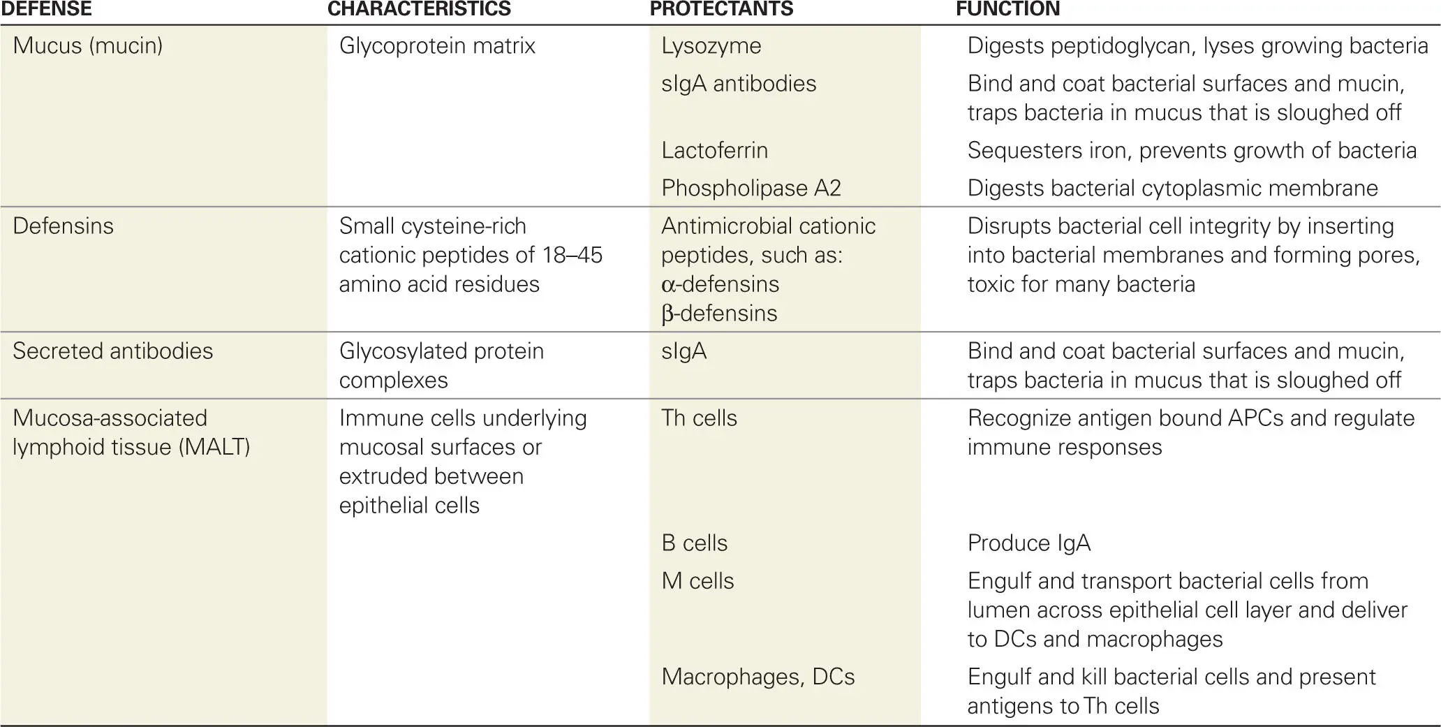
Table 2-3. Special defenses of specific sites
Normal Microbiota of the Skin and Mucosa
Читать дальшеИнтервал:
Закладка:
Похожие книги на «Bacterial Pathogenesis»
Представляем Вашему вниманию похожие книги на «Bacterial Pathogenesis» списком для выбора. Мы отобрали схожую по названию и смыслу литературу в надежде предоставить читателям больше вариантов отыскать новые, интересные, ещё непрочитанные произведения.


Обсуждение, отзывы о книге «Bacterial Pathogenesis» и просто собственные мнения читателей. Оставьте ваши комментарии, напишите, что Вы думаете о произведении, его смысле или главных героях. Укажите что конкретно понравилось, а что нет, и почему Вы так считаете.