Ceri Davies - Textbook for Orthodontic Therapists
Здесь есть возможность читать онлайн «Ceri Davies - Textbook for Orthodontic Therapists» — ознакомительный отрывок электронной книги совершенно бесплатно, а после прочтения отрывка купить полную версию. В некоторых случаях можно слушать аудио, скачать через торрент в формате fb2 и присутствует краткое содержание. Жанр: unrecognised, на английском языке. Описание произведения, (предисловие) а так же отзывы посетителей доступны на портале библиотеки ЛибКат.

- Название:Textbook for Orthodontic Therapists
- Автор:
- Жанр:
- Год:неизвестен
- ISBN:нет данных
- Рейтинг книги:4 / 5. Голосов: 1
-
Избранное:Добавить в избранное
- Отзывы:
-
Ваша оценка:
Textbook for Orthodontic Therapists: краткое содержание, описание и аннотация
Предлагаем к чтению аннотацию, описание, краткое содержание или предисловие (зависит от того, что написал сам автор книги «Textbook for Orthodontic Therapists»). Если вы не нашли необходимую информацию о книге — напишите в комментариях, мы постараемся отыскать её.
Written to help the reader understand the role and function of an orthodontic therapist,
offers support to those undertaking the Diploma in Orthodontic Therapy and to assist those who already work as orthodontic therapists, helping them in their quest to enhance safe and effective care.
Textbook for Orthodontic Therapists — читать онлайн ознакомительный отрывок
Ниже представлен текст книги, разбитый по страницам. Система сохранения места последней прочитанной страницы, позволяет с удобством читать онлайн бесплатно книгу «Textbook for Orthodontic Therapists», без необходимости каждый раз заново искать на чём Вы остановились. Поставьте закладку, и сможете в любой момент перейти на страницу, на которой закончили чтение.
Интервал:
Закладка:
2.2.1 Antero‐posterior Plane
The patient is viewed from the side.
This stage looks at the patient's skeletal pattern in the AP plane (front to back).
It assesses the patient’s profile and the relationship of the maxilla, referred to as the A point, and the mandible, referred to as the B point.
Patients are assessed by the following:Skeletal class I: Mandible is 2–3 mm posterior to the maxilla ( Figure 2.1).Skeletal class II: Mandible is retruded relative to the maxilla ( Figure 2.2).Skeletal class III: Mandible is protruded relative to the maxilla ( Figure 2.3).
2.2.2 Vertical Plane
The patient is viewed from the front and side.
This stage looks at the patient’s skeletal pattern in the vertical plane (up and down).
It assesses the lower part of the face, looking at the LAFH (lower anterior facial height) and FMPA (Frankfort‐mandibular plane angle).
LAFH:
LAFH should measure the same as UAFH (upper anterior facial height). Figure 2.1 Skeletal class I. Figure 2.2 Skeletal class II. Figure 2.3 Skeletal class III.
UAFH is measured from the eyebrow to the base of nose ( Figure 2.4).
LAFH is measured from the base of the nose to the lowest point on the chin.
FMPA:
This is the angle where the Frankfort plane and mandibular plane meet ( Figure 2.5).
This is assessed by placing one hand on the Frankfort plane and one hand on the mandibular plane and assessing where they cross by eye.
The measurements indicate the following:
Average LAFH and FMPA – usually seen in class I.
Decreased LAFH and low FMPA – usually seen in class II.
Increased LAFH and high FMPA – usually seen in class III.
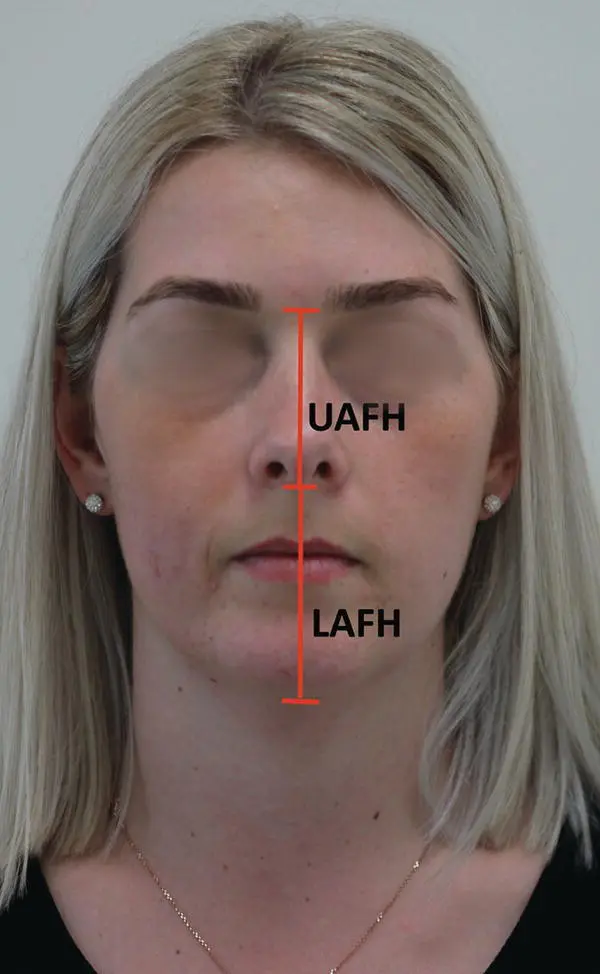
Figure 2.4 Vertical plane measurements: frontal view.
2.2.3 Transverse Plane
The patient is viewed from the front and looking down on the face from above.
This stage looks at the patient’s skeletal pattern in the transverse plane (side to side).
This is assessed by looking at the patient’s face and recording any significant asymmetry ( Figure 2.6) – all patients are asymmetrical. Figure 2.5 Vertical plane measurements: right profile. Figure 2.6 Transverse plane: frontal view looking for any asymmetry.
To assess whether the occlusal plane follows the line of asymmetry, a tongue spatula is used and the patient is asked to bite onto this ( Figure 2.7). In patients who have a significant asymmetry, this will be noticeable by the tongue spatula not lying flat.
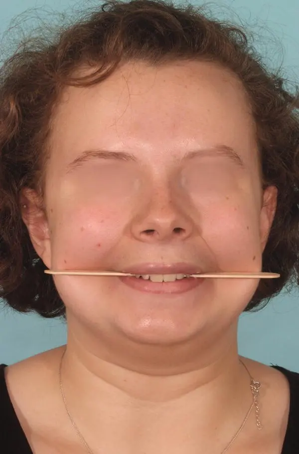
Figure 2.7 Transverse plane: frontal view looking for any asymmetry with a spatula.
2.2.4 Profile Pattern
This is assessed by looking at the LAFH.
Patients can present with different profiles: concave, straight, or convex ( Figure 2.8).
In the convex profile the LAFH is protruded, most commonly found in African Caribbean people.
In the straight profile the LAFH is straight from the nose to the chin.
In the concave profile the LAFH is caved in between the nose and the chin.
Patients with a concave profile should be approached with care when it comes to extractions, as this could make them even more concave, whereas convex profile patients would benefit from this.
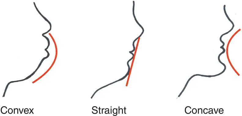
Figure 2.8 The different profile patterns.
2.3 Intra‐Oral Assessment
There are many stages considered when carrying out an intra‐oral assessment. The first three look at the three planes of space, whereas the remaining eight look more into how the dentition appears in the mouth.
2.3.1 Anterio‐posterior Plane
Assesses the arch from front to back.
This stage looks at the different types of relationships of the occlusion, such as molar relationship, incisor relationship, canine relationship, and overjet.
Molar relationship:Also known as Angle’s classification.Looks at the position of the upper first molars.Classification is class I; class II, II25, II50, II70; and class III, III25, III50, III70.
Incisor relationship:Also known as the BSIC (British Standards Institute Classification 1983).Looks at the position of the incisors.Classification is Class I; Class II div I, div II; and Class III.
Canine relationship:Looks at the position of the canines.Classification is class I; class II, II25 II50, II70; and class III, III25, III50, III70.
Overjet:Distance between the upper and lower teeth within the horizontal plane. The distance between them is measured with a stainless‐steel ruler.
2.3.2 Vertical Plane
Assesses the arch up and down.
Looks at the overbite.
The overbite is the vertical overlap of the upper incisors over the lower incisors when the posterior teeth are in occlusion. It can be measured as:Average: upper incisors cover one‐third of the lower incisors when in occlusion.Increased and complete: upper incisors cover more than one‐third of the lower incisors, but are complete (touching) on hard or soft tissues when in occlusion.Increased and incomplete: upper incisors cover more than one‐third of the lower incisors and are incomplete (not touching) on hard or soft tissues when in occlusion.Decreased and complete: upper incisors cover less than one‐third of the lower incisors, but are complete (touching) on hard or soft tissues when in occlusion.
Decreased and incomplete: upper incisors cover less than one‐third of the lower incisors and are incomplete (not touching) on hard or soft tissues when in occlusion.
2.3.3 Transverse Plane
Assesses the arch from side to side.
Looks for any posterior crossbites.
A posterior crossbite is found when the upper dentition on the posterior segment sits within the lower teeth when occluding.
There can be unilateral or bilateral crossbites, unilateral meaning on one side of the arch and bilateral being on both sides of the arch.
2.3.4 Crowding
This assesses the amount of crowding there is within both the upper and lower arches.
Assessing the crowding falls into the category of space analysis.
Constructing an archform that best fits the majority of the teeth can help analyse how much crowding there is within the arch.
Once the archform has been constructed, it is then measured by the use of floss and a stainless‐steel ruler. Once the measurement has been calculated for that, the mesiodistal widths of all the teeth within that arch are measured. The calculations are then taken away from each other, which gives you the total amount of crowding:
Crowding is then classified as the following:Mild crowding: 2–4 mmModerate crowding: 4–8 mmSevere crowding: 8+ mm.
2.3.5 Spacing
This assesses the amount of spacing that is present within the two arches.
Spacing can be classified into mild, moderate, or severe.
Spacing can be found within the arch due to the following:Extractions: patients who have previously had extractions, for example extracted LL6.Dento‐alveolar disproportion: patients who have big arches but small (microdontia) teeth can present with spacing between the teeth.Microdontia: patient who present with small teeth, for example peg laterals.Diastema: patients who present with a diastema within the arch, which could be due to a habit such as thumb sucking, active tongue thrust, or a mesiodens lying erupted or unerupted between the upper central incisors.
Читать дальшеИнтервал:
Закладка:
Похожие книги на «Textbook for Orthodontic Therapists»
Представляем Вашему вниманию похожие книги на «Textbook for Orthodontic Therapists» списком для выбора. Мы отобрали схожую по названию и смыслу литературу в надежде предоставить читателям больше вариантов отыскать новые, интересные, ещё непрочитанные произведения.


Обсуждение, отзывы о книге «Textbook for Orthodontic Therapists» и просто собственные мнения читателей. Оставьте ваши комментарии, напишите, что Вы думаете о произведении, его смысле или главных героях. Укажите что конкретно понравилось, а что нет, и почему Вы так считаете.