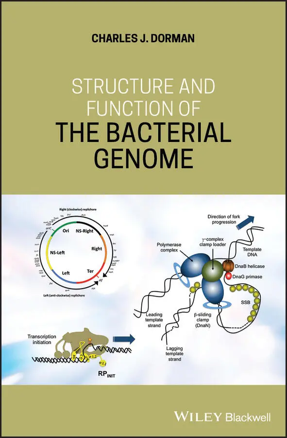
Charles J. Dorman - Structure and Function of the Bacterial Genome
Здесь есть возможность читать онлайн «Charles J. Dorman - Structure and Function of the Bacterial Genome» — ознакомительный отрывок электронной книги совершенно бесплатно, а после прочтения отрывка купить полную версию. В некоторых случаях можно слушать аудио, скачать через торрент в формате fb2 и присутствует краткое содержание. Жанр: unrecognised, на английском языке. Описание произведения, (предисловие) а так же отзывы посетителей доступны на портале библиотеки ЛибКат.
- Название:Structure and Function of the Bacterial Genome
- Автор:
- Жанр:
- Год:неизвестен
- ISBN:нет данных
- Рейтинг книги:5 / 5. Голосов: 1
-
Избранное:Добавить в избранное
- Отзывы:
-
Ваша оценка:
Structure and Function of the Bacterial Genome: краткое содержание, описание и аннотация
Предлагаем к чтению аннотацию, описание, краткое содержание или предисловие (зависит от того, что написал сам автор книги «Structure and Function of the Bacterial Genome»). Если вы не нашли необходимую информацию о книге — напишите в комментариях, мы постараемся отыскать её.
Structure and Function of the Bacterial Genome Blends knowledge of gene regulatory mechanisms with a consideration of nucleoid structure and dynamics Offers a 'DNA-centric' approach to considering transcription control Views horizontal gene transfer from a gene regulation perspective Assesses the opportunities and limitations of designing synthetic microbes or rewiring existing ones
is an ideal book for graduate and undergraduate students studying microbial cell biology, bacterial pathogenesis, gene regulation, and molecular microbiology. It will also appeal to principal investigators conducting research on these and related topics and researchers in synthetic biology and other arms of biotechnology.
Structure and Function of the Bacterial Genome — читать онлайн ознакомительный отрывок
Ниже представлен текст книги, разбитый по страницам. Система сохранения места последней прочитанной страницы, позволяет с удобством читать онлайн бесплатно книгу «Structure and Function of the Bacterial Genome», без необходимости каждый раз заново искать на чём Вы остановились. Поставьте закладку, и сможете в любой момент перейти на страницу, на которой закончили чтение.
Интервал:
Закладка:
Decatenation of the interlinked chromosome copies by topoisomerase IV and the resolution of any chromosome multimers by XerCD are needed prior to sister chromosome segregation (Hiraga 1993). In the absence of Topo IV, XerCD‐ dif‐ FtsK can achieve the same outcome by a process of local reconnection involving multiple rounds of site‐specific recombination (Grainge et al. 2007). Finally, the segregation process will move one chromosome, together with any associated live replication forks, into one of the daughter cells. In E. coli and its close relatives, this occurs without the aid of a dedicated protein‐based active partitioning system equivalent to the ParAB proteins and the parS cis ‐acting partitioning DNA sequence that are found in most other bacteria (Badrinarayanan et al. 2015; Bignell and Thomas 2001). Radial confinement of the two sister chromosomes has been proposed as playing a role in segregation in the case of E. coli . Here the two chromosome polymers repel one another through an entropic exclusion mechanism that drives the copies into separate compartments before the closure of the cell division septum (Jun and Wright 2010; Junier et al. 2014).
The specificity of chromosome orientation within the cytoplasm has led to the interesting proposal that the chromosome provides the prokaryotic cell with an internal frame of reference, something that has been lost in eukaryotes because the genetic material there is in a membrane‐enclosed nucleus (Theriot 2013). This reference frame is useful in providing each molecule in the cell with a set of spatial coordinates. Developing the point further, Theriot has proposed that eukaryotes rely on their cytoskeleton to provide a reference frame (Theriot 2013). We will return to the issue of spatial and temporal positioning of molecules ( Chapter 8).
1.20 SeqA and Nucleoid Organisation
DNA‐binding proteins play important roles in organising the structure of the folded chromosome, with some of these proteins having a chromosome‐domain‐specific binding pattern (Dame et al. 2011). The SeqA protein was introduced during the description of factors involved in the control of the initiation of chromosome replication ( Section 1.3). SeqA accompanies the moving replication fork (Brendler et al. 2000; Onogi et al. 1999), resulting in a dynamic pattern of binding around the chromosome (Sánchez‐Romero et al. 2010; Waldminghaus and Skarstad 2010). This protein seems to be excluded from binding within the Ter macrodomain, possibly reflecting the absence from Ter of matches to the consensus sequence for SeqA high‐affinity binding sites (Sánchez‐Romero et al. 2010; Waldminghaus and Skarstad 2010). The SeqA protein can interact with the cell envelope as well as hemimethylated DNA (D'Alençon et al. 1999; Mika et al. 2015; Shakibai et al. 1998; Slater et al. 1995) so may it play a role in the positioning of Ori during cell division (Dame et al. 2011).
1.21 MukB, a Condensin‐Like Protein
The bacterial chromosome is maintained in an orderly superstructure to facilitate replication, transcription, and other DNA‐based transactions. The family of SMC proteins play an important role in achieving this organisation (Uhlmann 2016). These large polypeptides have a DNA‐binding head domain and long coiled‐coil domains that bring the head‐domain‐DNA complexes together in a condensed nucleoprotein complex ( Figure 1.10). The head domains have ATPase activity and a DNA‐binding hinge region in the coiled‐coil domain promotes dimer formation (Chen, N., et al. 2008; Kumar et al. 2017a). SMC activity is found in eukaryotes and in prokaryotes, with one of the best‐studied examples of an SMC‐like protein in bacteria being the MukB protein from E. coli (Niki et al. 1991; Rybenkov et al. 2014).
MukB forms topologically stable loops in the chromosomal DNA and protects the supercoils in these protected loops (Kumar et al. 2017a). It forms a complex with the MukE and MukF proteins, with these seeming to play a role in the turnover of the MukB complex on DNA in combination with the ATPase activity of MukB itself (Kumar et al. 2017a). MukF performs a bridging role between the ATPase heads of the two MukB in the complex ( Figure 1.10). Proteins performing this task in SMC complexes are called kleisins. The equivalent system in B. subtilis and C. crescentus consists of the proteins Smc (MukB), ScpA (MukF, kleisin), and ScpB (MukE): the ‘Scp’ designation indicates that the protein is a ‘segregation and condensation protein’ (Britton et al. 1998; Burmann et al. 2013; Jensen and Shapiro 1999, 2003; Mascarenhas et al. 2005).
The MukBEF complex has important architectural and segregational roles in the nucleoid, operating mainly outside the Ter macrodomain of the chromosome. Its principal site of action seems to be at ori and MukB requires MukE, MukF, and ATP hydrolysis to gain and maintain this association; MukBEF/SMC complexes do not seem to track moving replisomes (Badrinarayanan et al. 2012a,b; Danilova et al. 2007; Gruber and Errington 2009; Sullivan et al. 2009). MukBEF is responsible for guiding the newly replicated ori regions into the two halves of the cell, driving bipolar segregation; if MukBEF is removed, the ori shifts from mid‐cell to the pole, disturbing normal chromosome orientation and segregation patterns (Danilova et al. 2007). Inside the Ter macrodomain, the MatP protein prevents MukBEF from playing a structural role by displacing it and so making it available for ori binding (Lioy et al. 2018; Nolivos and Sherratt 2014; Nolivos et al. 2016). The MukBEF complex is not required for sister chromosome cohesion because muk mutants have a higher degree of cohesion of sister chromosomes than wild‐type cells (Danilova et al. 2007).
Despite being able to stimulate Topo IV activity by direct interaction (Hayama and Marians 2010; Hayama et al. 2013; Li et al. 2010; Vos et al. 2013), MukBEF forms a complex with Topo IV that seems to stabilise MukBEF on the DNA and drive chromosome condensation independently of the catalytic activities of the topoisomerase (Kumar et al. 2017b). This observation is indicative of an architectural role for the MukBEF‐Topo‐IV complex. Data from chromosome conformation capture experiments show that, together with the HU NAP, MukBEF promotes long‐range DNA contacts in the megabase range (Lioy et al. 2018), adding further detail to our picture of the nucleoid‐structuring contributions of MukBEF. However, the MukBEF‐Topo‐IV complex also has functions in chromosome segregation that do depend on the catalytic properties of Topo IV. The complex is responsible for the timely decatenation of newly replicated ori copies and their segregation ( Figure 1.10), while also contributing to the management of DNA supercoiling set points (Zawadzki et al. 2015). The primary site of action of the MukBEF‐Topo‐IV complex has been regarded as ori , with MukBEF recruiting the topoisomerase to that region of the chromosome (Nicolas et al. 2014). It is becoming clear that MukBEF‐Topo‐IV has a much more dynamic relationship with the chromosome, both spatially and temporally, and that its relationship with the MatP protein in ter has an important management role in its choreography. The final stages of chromosome replication and the associated need to effect decatenation in a timely manner are consistent with a need to grant MukBEF‐Topo‐IV access to ter to perform the necessary cohesion/decatenation steps, with MatP then evicting the complex, making it available for re‐association with ori (Nolivos et al. 2016).
1.22 MatP, the matS Site and Ter Organisation
The 17‐kDa MatP protein binds to 23 high‐affinity matS sites found exclusively within the Ter macrodomain of the E. coli chromosome (Mercier et al. 2008; Thiel et al. 2012). The Ter domains of both chromosome I and chromosome II of V. cholerae also have matS sites that are bound by MatP and these affect the spatiotemporal coordination of the replication and segregation of both chromosomes (Demarre et al. 2014). Counterparts of matS are present in the Ter regions of the chromosomes of Erwinia carotovora , S. enterica serovar Typhimurium LT2, and Yersinia pestis (Mercier et al. 2008). MatP forms bridged tetramers that link distant matS sites to condense the Ter DNA (Dupaigne et al. 2012). MatP interacts with ZapB, a cell division‐associated protein, to position the Ter macrodomain at the cell midpoint and to ensure its segregation (Espéli et al. 2012). With proteins ZapA, C, and D, ZapB functions to organise the Z ring that acts as a scaffold for the assembly of the complexes responsible for cell division (Buss et al. 2013). ZapB is dependent on ZapA for interaction with the tubulin‐like FtsZ protein (Galli and Gerdes 2010). MatP also interacts with the MukBEF‐Topo‐IV complex at the Ter macrodomain, displacing MukBEF from Ter and facilitating its interaction with the Ori macrodomain. This is part of a step‐wise process in which MukBEF manoeuvres the chromosome such that Ori and Ter adopt their assigned positions at the pole and mid‐cell, respectively, by the time cell division takes place (Lioy et al. 2018; Nolivos et al. 2016). Together with MukBEF and SeqA, MatP is part of a group of proteins found exclusively in bacteria that express the Dam methyltransferase (Brezellec et al. 2006; Mercier et al. 2008).
Читать дальшеИнтервал:
Закладка:
Похожие книги на «Structure and Function of the Bacterial Genome»
Представляем Вашему вниманию похожие книги на «Structure and Function of the Bacterial Genome» списком для выбора. Мы отобрали схожую по названию и смыслу литературу в надежде предоставить читателям больше вариантов отыскать новые, интересные, ещё непрочитанные произведения.


Обсуждение, отзывы о книге «Structure and Function of the Bacterial Genome» и просто собственные мнения читателей. Оставьте ваши комментарии, напишите, что Вы думаете о произведении, его смысле или главных героях. Укажите что конкретно понравилось, а что нет, и почему Вы так считаете.