Tina M. Henkin - Snyder and Champness Molecular Genetics of Bacteria
Здесь есть возможность читать онлайн «Tina M. Henkin - Snyder and Champness Molecular Genetics of Bacteria» — ознакомительный отрывок электронной книги совершенно бесплатно, а после прочтения отрывка купить полную версию. В некоторых случаях можно слушать аудио, скачать через торрент в формате fb2 и присутствует краткое содержание. Жанр: unrecognised, на английском языке. Описание произведения, (предисловие) а так же отзывы посетителей доступны на портале библиотеки ЛибКат.
- Название:Snyder and Champness Molecular Genetics of Bacteria
- Автор:
- Жанр:
- Год:неизвестен
- ISBN:нет данных
- Рейтинг книги:3 / 5. Голосов: 1
-
Избранное:Добавить в избранное
- Отзывы:
-
Ваша оценка:
Snyder and Champness Molecular Genetics of Bacteria: краткое содержание, описание и аннотация
Предлагаем к чтению аннотацию, описание, краткое содержание или предисловие (зависит от того, что написал сам автор книги «Snyder and Champness Molecular Genetics of Bacteria»). Если вы не нашли необходимую информацию о книге — напишите в комментариях, мы постараемся отыскать её.
Although the text is centered on the most-studied bacteria,
and
, many examples are drawn from other bacteria of experimental, medical, ecological, and biotechnological importance. The book's many useful features include
Text boxes to help students make connections to relevant topics related to other organisms, including humans A summary of main points at the end of each chapter Questions for discussion and independent thought A list of suggested readings for background and further investigation in each chapter Fully illustrated with detailed diagrams and photos in full color A glossary of terms highlighted in the text While intended as an undergraduate or beginning graduate textbook, Molecular Genetics of Bacteria is an invaluable reference for anyone working in the fields of microbiology, genetics, biochemistry, bioengineering, medicine, molecular biology, and biotechnology.
"This is a marvelous textbook that is completely up-to-date and comprehensive, but not overwhelming. The clear prose and excellent figures make it ideal for use in teaching bacterial molecular genetics."—
, University of Washington
Snyder and Champness Molecular Genetics of Bacteria — читать онлайн ознакомительный отрывок
Ниже представлен текст книги, разбитый по страницам. Система сохранения места последней прочитанной страницы, позволяет с удобством читать онлайн бесплатно книгу «Snyder and Champness Molecular Genetics of Bacteria», без необходимости каждый раз заново искать на чём Вы остановились. Поставьте закладку, и сможете в любой момент перейти на страницу, на которой закончили чтение.
Интервал:
Закладка:
Some bacteria that live in and on our bodies also benefit us directly. The role of our commensal bacteria in human health is only beginning to be appreciated. It has been estimated that of the 10 14cells in a human body, only half are human! Of course, bacterial cells are much smaller than our cells, but this shows how our bodies are adapted to live with an extensive bacterial microbiome, which helps us digest food and avoid disease, among other roles, many of which are yet to be uncovered.
Bacteria have also long been used to make many useful compounds, such as antibiotics, and chemicals, such as benzene and citric acid. Bacteria and their bacteriophages are also the source of many of the useful enzymes used in molecular biology.
In spite of substantial progress, we have only begun to understand the bacterial world around us. Bacteria are the most physiologically diverse organisms on Earth, and the importance of bacteria to life on Earth and the potential uses to which bacteria can be put can only be guessed. Thousands of different types of bacteria are known, and new insights into their cellular mechanisms and their applications constantly emerge from research with bacteria. Moreover, it is estimated that less than 1% of the types of bacteria living in the soil and other environments have ever been isolated. Recent culture-independent mechanisms indicate that bacterial diversity is much greater than we ever imagined (see Hug et al., Suggested Reading). In this new picture, it seems that less than half of the major lineages of bacteria have representatives that have been cultured. Organisms in these uncharacterized groups of bacteria may have all manner of interesting and useful functions. Clearly, studies of bacteria will continue to be essential to our future efforts to understand, control, and benefit from the biological world around us, and bacterial molecular genetics will be an essential tool in these efforts. However, before discussing this field, we must first briefly discuss the evolutionary relationship of bacteria to other organisms.
The Biological Universe
The Bacteria
This textbook comes at a very exciting time in our understanding of the interrelationship of all living things on the planet. After the landmark work of Carl Woese, all organisms on Earth were assigned to three major groups called domains: the bacteria (formerly eubacteria), the archaea (formerly archaebacteria), and the eukaryotes (see Woese and Fox, Suggested Reading). However, it is now clear that two major divisions account for these three groups. Bacteria form one of these divisions, while eukaryotes are now believed to have diverged out of the archaea. Figure 1shows the microbiologists’ view of the living world, where microbes provide most of the diversity and eukaryotes occupy a relatively small niche. This is not a far-fetched concept. Sequence data show that we differ from chimpanzees by only 2% of our DNA sequence, while 25 to 50% of the genes in a typical bacterium are unique to the species. Furthermore, while mammals diverged from each other on the order of millions of years ago, the main bacterial lineages diverged billions of years ago.
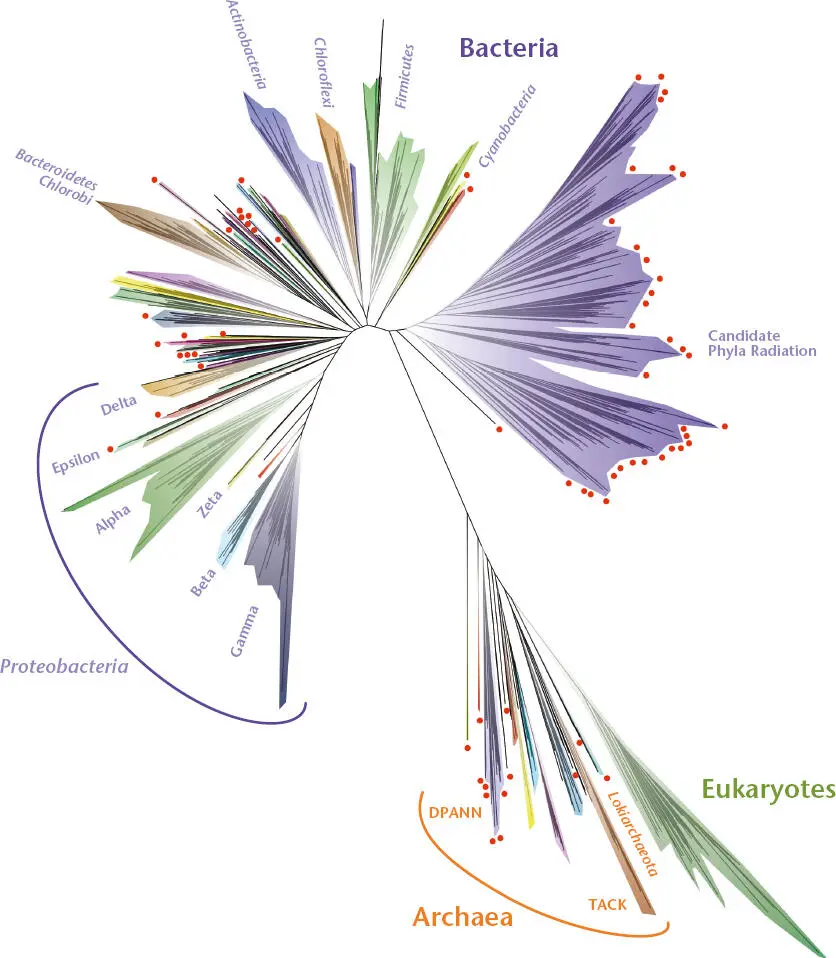
Figure 1 A molecular tree of life capturing diversity using ribosomal proteins from sequenced genomes (see Hug et al., Suggested Reading). Selected major linages within bacteria are indicated, including the Proteobacteria and the subgroups Alpha, Beta, Delta, Epsilon, Gamma, and Zeta, the Firmicutes , and the Candidate Phyla Radiation, which is almost completely devoid of cultured representatives. For the Archaea , two superphyla, TACK and DPANN, are indicated and described in the text. The position of the archaeal Lokiarchaeota lineage is indicated. Genome sequences from members of the Lokiarchaeota lineage indicate that they possess molecular systems previously believed to be found only in Eukaryotes . Red dots indicate lineages that have no cultured representatives. Adapted with permission from Hug L, et al, Nat Microbiol 1:16048 (2016), https://doi.org/10.1038/nmicrobiol.2016.48.
Bacteria can differ greatly in their physical appearance under the microscope. Although most are single celled and rod shaped or spherical, some are multicellular and undergo complicated developmental cycles. The cyanobacteria (formerly called blue-green algae) are bacteria, but they have chlorophyll and can be filamentous, which is why they were originally mistaken for algae. The antibiotic-producing actinomycetes, which include Streptomyces spp., are also bacteria, but they form hyphae and stalks of spores, making them resemble fungi. Another bacterial group, the Caulobacter spp., have both free-swimming and sessile forms that attach to surfaces through a holdfast structure. Some of the most dramaticappearing bacteria of all belong to the genus Myxococcus , members of which can exist as free-living single-celled organisms but can also aggregate to form fruiting bodies, much like slime molds. As mentioned above, bacterial cells are usually much smaller than the cells of higher organisms, but one very large bacterium, Epulopiscium , can be over half a millimeter long, longer than even most eukaryotic cells (see Angert, Suggested Reading). In addition, unlike most bacteria that multiply by simple division, Epulopiscium gives birth to multiple live progeny. Despite the fact that some bacteria are found in dramatically different shapes and sizes, they cannot be distinguished simply by their physical appearance; instead, it is necessary to use biochemical criteria, such as the sequences of their ribosomal proteins or RNAs (rRNAs), whose sequences are characteristic of the three domains of life.
GRAM-NEGATIVE AND GRAM-POSITIVE BACTERIA
Bacteria have historically been divided into two major subgroups, the Gram-negativeand Gram-positivebacteria. This division was based on the response to a test called the Gram stain. “Gram-negative” bacteria retain little of the dye and are pink after this staining procedure, whereas “Gram-positive” bacteria retain more of the dye and turn deep blue. The difference in staining typically reflects the fact that Gram-negative bacteria are surrounded by a thinner structure composed of both an inner and an outer membrane, while the structure surrounding Gram-positive bacteria is much thicker, consisting of a single membrane surrounded by a thicker wall. However, this older form of classification is being replaced by talking about the phyla of bacteria as determined by the DNA sequence. The Firmicutes are a broad group containing Bacillus , clostridia, lactic acid bacteria, and the Tenericutes , including the mycoplasmas. Firmicutes have been referred to as low G+C Gram-positive bacteria based on the low percentage of guanine and cytosine (low G+C) compared to adenine and thymine often found in the genome sequence of members of this group (see chapter 1). However, having a low G+C genome is not a universal feature of the Firmicutes , which limits the utility of the designation. Another group of bacteria that were classically described as high G+C and Gram positive because they typically possess a higher percentage of guanine and cytosine includes the Actinobacteria (actinomycetes), such as Streptomyces and Mycobacterium .
The Gram designation system of classifying bacteria is particularly weak for capturing the diversity of the numerous phyla that stain Gram negative. While many bacteria historically referred to as Gram negative, such as Escherichia coli , Pseudomonas , and Rhizobium , fall within a broad group known as the Proteobacteria , many other characterized and uncharacterized groups also exist. It is also worth pointing out that relying on a staining form of classification is particularly contrived when talking about uncultured bacteria or those that are only capable of growth as symbionts in other organisms. Given all of these considerations, instead of using the Gram-positive and Gram-negative designations as a tool for describing the relatedness of groups of bacteria, we now opt for a more precise designation by referring to the phyla. In addition, while we will remind the reader of the older designations of Gram positive and Gram negative in the text when referring to the superstructure surrounding the cell, we have transitioned to describing the cell wall structure more directly.
Читать дальшеИнтервал:
Закладка:
Похожие книги на «Snyder and Champness Molecular Genetics of Bacteria»
Представляем Вашему вниманию похожие книги на «Snyder and Champness Molecular Genetics of Bacteria» списком для выбора. Мы отобрали схожую по названию и смыслу литературу в надежде предоставить читателям больше вариантов отыскать новые, интересные, ещё непрочитанные произведения.


Обсуждение, отзывы о книге «Snyder and Champness Molecular Genetics of Bacteria» и просто собственные мнения читателей. Оставьте ваши комментарии, напишите, что Вы думаете о произведении, его смысле или главных героях. Укажите что конкретно понравилось, а что нет, и почему Вы так считаете.