The Peripheral T-Cell Lymphomas
Здесь есть возможность читать онлайн «The Peripheral T-Cell Lymphomas» — ознакомительный отрывок электронной книги совершенно бесплатно, а после прочтения отрывка купить полную версию. В некоторых случаях можно слушать аудио, скачать через торрент в формате fb2 и присутствует краткое содержание. Жанр: unrecognised, на английском языке. Описание произведения, (предисловие) а так же отзывы посетителей доступны на портале библиотеки ЛибКат.
- Название:The Peripheral T-Cell Lymphomas
- Автор:
- Жанр:
- Год:неизвестен
- ISBN:нет данных
- Рейтинг книги:3 / 5. Голосов: 1
-
Избранное:Добавить в избранное
- Отзывы:
-
Ваша оценка:
The Peripheral T-Cell Lymphomas: краткое содержание, описание и аннотация
Предлагаем к чтению аннотацию, описание, краткое содержание или предисловие (зависит от того, что написал сам автор книги «The Peripheral T-Cell Lymphomas»). Если вы не нашли необходимую информацию о книге — напишите в комментариях, мы постараемся отыскать её.
The first book of its kind,
presents a far-reaching survey of this complex and rare group of blood cancers. Featuring contributions from thought-leaders concerned with all aspects of PTCL, this authoritative text covers biology, epidemiology, classification, approved and emerging drugs, molecular genetics, and more. Detailed clinical chapters address diagnosis, prognosis, and treatment of each of the major PTCL subtypes identified in the 2018 WHO Classification of Tumors of Hematopoietic and Lymphoid Tissues. This much-needed resource:
Covers the biological basis, epidemiology, classification, and treatment of PTCL Discusses the future of the field, including global collaboration efforts and novel approaches to PCTL Explores the role of biologics in PTCL and autologous and allogeneic stem-cell transplantation Offers new insights on molecular pathogenesis, innovative therapeutics, and novel drug combinations Features contributions from the Chairs The T-Cell Lymphoma Forum: the world’s largest meeting focused on PTCL Reflecting the unique epidemiology and genetic diversity of the PTCL,
is an indispensable source of data, insight, and references for the medical community, particularly those working in oncology and hematology.
The Peripheral T-Cell Lymphomas — читать онлайн ознакомительный отрывок
Ниже представлен текст книги, разбитый по страницам. Система сохранения места последней прочитанной страницы, позволяет с удобством читать онлайн бесплатно книгу «The Peripheral T-Cell Lymphomas», без необходимости каждый раз заново искать на чём Вы остановились. Поставьте закладку, и сможете в любой момент перейти на страницу, на которой закончили чтение.
Интервал:
Закладка:
More recently, increasing numbers of ALCL developing at the contact of textured breast implants have been reported. The disease presents most often with neoplastic T cells restricted to the seroma and/or the surface of the periprosthetic capsule (“in‐situ form”), and more rarely as a tumor mass within the capsule and the adjacent tissues (“infiltrative form”). While the infiltrative form of the disease can have an aggressive course and requires systemic chemotherapy, the in‐situ form usually regresses after capsulectomy. A model of lymphomagenesis induced by local inflammation and chronic antigenic stimulation is proposed. The resulting high levels of cytokines in the periprosthetic confined milieu could favor the expansion of cytotoxic cells, which would subsequently acquire somatic mutations. Several studies have identified recurrent mutations in epigenetic modifiers ( KMT2C, KMT2D, CHD2, CREBBP ) and JAK–STAT signaling ( STAT3, JAK1, STAT5B ) pathway) [28, 104]. The role of a bacteria, Ralstonia pickettii , in promoting inflammation, has been suspected, although this remains controversial [105].
Other Factors
Only few physical factors have been identified as underlying factors favoring T‐cell lymphoma development. Analyses of mutational signatures derived from whole genome sequencing revealed that enrichment in ultraviolet radiation‐induced signature in CTCL are, suggesting that ultraviolet radiation could contribute to CTCL [106].
The role of mutations in epigenetic regulators which occur at high frequency in hematopoietic progenitor cells (see section 2.2.1.1 Signaling Pathways) is still unclear. Beside changes in the gene expression levels induced by epigenetic alterations in the regulatory regions, these mutations could induce some genomic instability, subsequently favoring the acquisition of additional mutations that could drive the tumor transformation. It has been recently postulated that TET2, IDH2 , and DNMT3A could induce some degrees of genomic instability by various pathway. Mutations in IDH1/2 could also result, via the production of the oncometabolite D‐2 hydroxyglutarate, in impaired DNA repair, which could have therapeutic consequences [107, 108]. Alterations in TET proteins could also increase DNA damage [109, 110].
Few predisposing factors of PTCL development have been identified in population studies. In addition to HLA‐DPB1 association with NKTCL which could explain geographic differences in NKTCL incidence (see above), a germline HAVCR2 mutation altering TIM‐3 has been reported in around 60% of patients with subcutaneous panniculitis‐like T‐cell lymphoma [111].
Cell of Origin ( Table 2.1)
Cellular origin is a major defining feature of cancers in general. Likewise, normal counterpart is one of the defining features of lymphomas in the World Health Organization classification, and non‐Hodgkin lymphoma entities are primarily stratified according to their cellular derivation from B cells compared with T or NK cells.
In the B‐cell lineage, stages of normal B‐cell differentiation proceed through successive steps that occur in distinct microenvironments, are associated with genetic events, modification of gene expression profiles and immunophenotypes and to some extent morphology, and there is a relatively good correspondence of many B‐cell neoplasms to normal counterparts [112].
In contrast, the cellular immune system comprises multiple functional subsets featuring functional plasticity, and NK/T‐cell activation and differentiation do not encompass acquired genetic traits. One important level of dichotomy is between the innate and the acquired immune systems which comprise distinct cellular subsets with different physiological properties, and the derived neoplasms broadly feature different characteristics, emphasizing the concept that cellular derivation is an important determinant of lymphoma biology ( Figure 2.3). Innate immune cells comprise NK cells, γδ T cells and a minor subset of αβ T cells. These cells preferentially distributed at the interface with external antigenic stimuli (skin and mucosae) provide immediate immune responses which are not restricted by antigen recognition in the context of MHC presentation, and lack immunological memory. NK cells lack TCR gene rearrangements and lack membrane TCR expression. NK cells share some markers with T cells such as CD2, CD7, CD45RO, and cytoplasmic (but not surface) CD3. NK cells are usually CD4–/CD8– but may be CD8+, and they express one or several of the “NK‐associated” antigens (CD11b, CD16, CD56, CD57, NK receptors), which are, however, not entirely specific. γδ T cells (CD4‐CD8‐ or more rarely CD4–/CD8+) comprise less than 5% of T cells and are preferentially distributed in the skin, mucosae, and to the splenic red pulp. Both NK cells and γδ T cells express cytotoxic proteins, T‐cell intracellular antigen‐1 (a marker of cytotoxic cells in general), perforin and granzyme B (both expressed upon activation and not in the resting stage). The majority of T lymphocytes are αβ T cells, which recognize the antigen in a MHC‐restricted fashion in the presence of an antigen‐presenting cell, and constitute the adaptative immune system, which is characterized by specificity and memory of the immunological response.
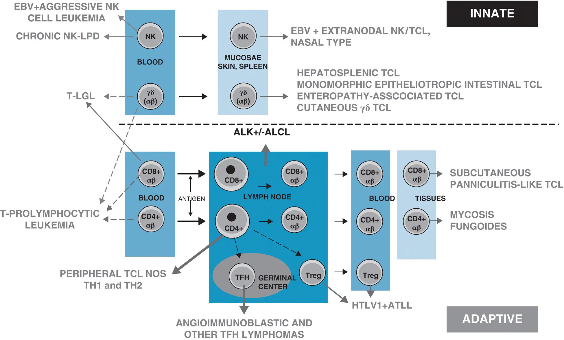
Figure 2.3 Cellular derivation of T‐cell and NK‐cell lymphomas from the innate and adaptive immune systems. ALCL, anaplastic large‐cell lymphoma; ALK, anaplastic lymphoma kinase; EBV, Epstein–Barr virus; LPD, lymphoproliferative disorders; NK, natural killer; TCL, T‐cell lymphoma; TFH, T follicular helper cells; TH1, 2, T lymphocyte 1, 2; Treg, T‐regulatory cells.
Source: Adapted from de Leval and Gaulard [122].
Neoplasms derived from the innate immune system comprise several mostly extranodal cytotoxic malignancies, which are overall rare and, with the exception of the usually indolent chronic leukemias of NK cells or large granular lymphocytes, often have a highly aggressive behavior and poor outcome. Innate‐type lymphomas may occur in young individuals and some entities, such as ENKTL, are defined by their association to EBV infection. Together with the cytotoxic features of the malignant cells and/or EBV infection, these diseases may not uncommonly be associated to necrosis and a hemophagocytic syndrome [113]. Although there might be a predilection for derivation from NK compared with γδ compared with αβ T cells in distinct entities, only the NK‐cell leukemias and primary cutaneous γδ T‐cell lymphomas are by definition homogeneous in terms of cellular lineage. Interestingly, there is marked overlap in the mutational landscapes of these different innate lymphoid neoplasms. Mutation‐induced activation of the JAK–STAT pathway is highly recurrent among those [114], and inactivation of the SETD2 histone methyltransferase appears to be tightly linked to entities derived from gamma‐delta T cells, MEITL, and HSTL [23, 54, 114].
It is also remarkable that extranodal PTCL entities tend to have a predilection for development in peculiar organs. Accordingly, distinct extranodal PTCL entities have a gene expression signature component that is organ specific [115, 116]. In some instances, the association reflects derivation from a subset of organ‐specific lymphocytes, or lymphocytes with peculiar homing properties. For example, EATL and MEITL are derived from intraepithelial lymphocytes of the intestinal mucosa. Most cases of HSTL derive from functionally immature cytotoxic γδ T cells of the splenic pool with Vδ1 gene usage. Epidermotropic mycosis fungoides, which is the most common cutaneous lymphoma, is associated with an expansion of lymphoid cells with homing properties to the skin. Altogether, this suggests the importance of tissue‐specific factors in sustaining or promoting tumor growth. Interestingly, a feature common to extranodal PTCL entities is the rarity of dissemination to the bone marrow and the lymph nodes.
Читать дальшеИнтервал:
Закладка:
Похожие книги на «The Peripheral T-Cell Lymphomas»
Представляем Вашему вниманию похожие книги на «The Peripheral T-Cell Lymphomas» списком для выбора. Мы отобрали схожую по названию и смыслу литературу в надежде предоставить читателям больше вариантов отыскать новые, интересные, ещё непрочитанные произведения.


Обсуждение, отзывы о книге «The Peripheral T-Cell Lymphomas» и просто собственные мнения читателей. Оставьте ваши комментарии, напишите, что Вы думаете о произведении, его смысле или главных героях. Укажите что конкретно понравилось, а что нет, и почему Вы так считаете.