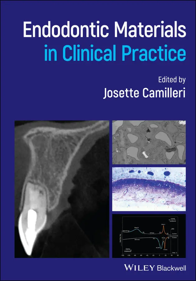
Endodontic Materials in Clinical Practice
Здесь есть возможность читать онлайн «Endodontic Materials in Clinical Practice» — ознакомительный отрывок электронной книги совершенно бесплатно, а после прочтения отрывка купить полную версию. В некоторых случаях можно слушать аудио, скачать через торрент в формате fb2 и присутствует краткое содержание. Жанр: unrecognised, на английском языке. Описание произведения, (предисловие) а так же отзывы посетителей доступны на портале библиотеки ЛибКат.
- Название:Endodontic Materials in Clinical Practice
- Автор:
- Жанр:
- Год:неизвестен
- ISBN:нет данных
- Рейтинг книги:4 / 5. Голосов: 1
-
Избранное:Добавить в избранное
- Отзывы:
-
Ваша оценка:
Endodontic Materials in Clinical Practice: краткое содержание, описание и аннотация
Предлагаем к чтению аннотацию, описание, краткое содержание или предисловие (зависит от того, что написал сам автор книги «Endodontic Materials in Clinical Practice»). Если вы не нашли необходимую информацию о книге — напишите в комментариях, мы постараемся отыскать её.
Presents an illustrated guide to all materials used in endodontic practice Focuses on the clinical application for each material Explains why specific materials are used Includes information on how to select the correct material Considers locations and techniques in making material decisions Written for specialist endodontists and residents, dental material specialists, post-graduate students, general dentists, and dentistry students,
is an essential resource for selecting the right materials for specific techniques.
Endodontic Materials in Clinical Practice — читать онлайн ознакомительный отрывок
Ниже представлен текст книги, разбитый по страницам. Система сохранения места последней прочитанной страницы, позволяет с удобством читать онлайн бесплатно книгу «Endodontic Materials in Clinical Practice», без необходимости каждый раз заново искать на чём Вы остановились. Поставьте закладку, и сможете в любой момент перейти на страницу, на которой закончили чтение.
Интервал:
Закладка:
6 Chapter 6Figure 6.1 Penetration of a sealer, dyed with Sudan Black B, at different le...Figure 6.2 Confocal microscope image (×10) of penetration of AH Plus (Dentsp...Figure 6.3 Confocal microscope image (×10) of penetration of MTA Fillapex (A...Figure 6.4 Mandibular left first molar root filled with silver points.Figure 6.5 Gutta‐percha cones of different sizes and tapers.Figure 6.6 Suboptimal, poorly condensed gutta‐percha root filling in a maxil...Figure 6.7 Extrusion of sealer from the mesiolingual canal of a mandibular r...Figure 6.8 Root canal sealer extruded through to reveal a lateral canal in a...Figure 6.9 Cold lateral condensation. (a) Spreader inserted and force used t...Figure 6.10 Maxillary left central incisor obturated using the single‐cone t...Figure 6.11 Continuous wave of condensation. (a) Down‐pack, heat, and vertic...Figure 6.12 Elements Free obturation unit (Kerr Endodontics).Figure 6.13 Gutta‐Condensor (Dentsply Maillefer).Figure 6.14 Malformed mandibular left second premolar. (a) Root canal treatm...Figure 6.15 Disposable MTA carriers with different tip sizes.
7 Chapter 7Figure 7.1 Flap outline showing (a) sulcular triangular flap with a mesial v...Figure 7.2 (a) Raised submarginal flap and buccal bone perforation around th...Figure 7.3 Details of an apical surgical procedure following incision and ra...Figure 7.4 Root‐end resection, preparation, and filling. (a) Resected root e...Figure 7.5 Clinical case showing the procedure for root perforation repair. ...Figure 7.6 Presentation of (a) IRM Figure 7.7 Various presentations of glass ionomer cement. (a) Fuji IX....Figure 7.8 Scanning electron micrograph of hydrated MTA, showing the microst...Figure 7.9 Scanning electron micrograph of hydrated MTA, showing porosity re...Figure 7.10 In‐house manufacturing process of MTA Angelus, showing the furna...Figure 7.11 (a) ProRoot MTA (Dentsply).Figure 7.12 Various MTA carrier systems developed to aid in dispensing mater...Figure 7.13 Characterization of BC‐RRM, showing the material microstructure....Figure 7.14 Presentation of BC‐RRM (Totalfill; FKG Dentaire, Switzerland): (...
8 Chapter 8Figure 8.1 Treatment decision tree for deciduous teeth with deep carious les...Figure 8.2 Pulpotomy technique. (a, b) Radiographic and clinical presentatio...Figure 8.3 Indirect pulp treatment in two deciduous molars of a healthy 5.5‐...Figure 8.4 Pulpotomy of deciduous molars with various HCSCs: radiographic pr...Figure 8.5 Primary molar root canal morphology from microcomputed tomography...Figure 8.6 Pulpectomy of a left mandibular second molar with ZOE. Healthy fi...Figure 8.7 Pulpectomy of a right mandibular second molar with ZOE. Healthy f...Figure 8.8 Pulpectomy of a left mandibular second molar with Metapex (Meta B...Figure 8.9 Pulpectomy of a left upper second molar with Endoflas (Sanlor & C...
9 Chapter 9Figure 9.1 Image made with polarized light, showing the presence of dentinal...Figure 9.2 Detail of root dentine, showing its translucent appearance in the...Figure 9.3 (a) Occlusal cavity with irregularities that disturb the chewing ...Figure 9.4 (a) Extensive restorations on teeth 14 and 15 with adhesive failu...
Guide
1 Cover Page
2 Title Page
3 Copyright Page
4 List of Contributors
5 Table of Contents
6 Begin Reading
7 Index
8 WILEY END USER LICENSE AGREEMENT
Pages
1 iii
2 iv
3 xi
4 xii
5 1
6 2
7 3
8 4
9 5
10 6
11 7
12 8
13 9
14 10
15 11
16 12
17 13
18 15
19 16
20 17
21 18
22 19
23 20
24 21
25 22
26 23
27 24
28 25
29 26
30 27
31 28
32 29
33 30
34 31
35 32
36 33
37 34
38 35
39 36
40 37
41 38
42 39
43 40
44 41
45 42
46 43
47 44
48 45
49 47
50 48
51 49
52 50
53 51
54 52
55 53
56 54
57 55
58 56
59 57
60 58
61 59
62 60
63 61
64 62
65 63
66 64
67 65
68 66
69 67
70 68
71 69
72 70
73 71
74 72
75 73
76 74
77 75
78 76
79 77
80 78
81 79
82 81
83 82
84 83
85 84
86 85
87 86
88 87
89 88
90 89
91 90
92 91
93 92
94 93
95 94
96 95
97 96
98 97
99 98
100 99
101 100
102 101
103 102
104 103
105 104
106 105
107 106
108 107
109 108
110 109
111 110
112 111
113 112
114 113
115 114
116 115
117 116
118 117
119 118
120 119
121 120
122 121
123 122
124 123
125 124
126 125
127 126
128 127
129 128
130 129
131 130
132 131
133 133
134 134
135 135
136 136
137 137
138 138
139 139
140 140
141 141
142 142
143 143
144 144
145 145
146 146
147 147
148 148
149 149
150 150
151 151
152 152
153 153
154 154
155 155
156 156
157 157
158 158
159 159
160 160
161 161
162 162
163 163
164 164
165 165
166 166
167 167
168 168
169 169
170 170
171 171
172 172
173 173
174 174
175 175
176 176
177 177
178 178
179 179
180 180
181 181
182 182
183 183
184 184
185 185
186 186
187 187
188 188
189 189
190 190
191 191
192 192
193 193
194 194
195 195
196 196
197 197
198 198
199 199
200 200
201 201
202 202
203 203
204 204
205 205
206 206
207 207
208 208
209 209
210 210
211 211
212 212
213 213
214 214
215 215
216 216
217 217
218 219
219 220
220 221
221 222
222 223
223 224
224 225
225 226
226 227
227 228
228 229
229 230
230 231
231 232
232 233
233 234
234 235
235 236
236 237
237 238
238 239
239 240
240 241
241 242
242 243
243 244
244 245
245 246
246 247
247 248
248 249
249 250
250 251
251 252
252 253
253 254
254 255
255 256
256 257
257 258
258 259
259 260
260 261
261 263
262 264
263 265
264 266
265 267
266 268
267 269
268 270
269 271
270 272
271 273
272 274
273 275
274 276
275 277
276 278
277 279
278 280
279 281
280 282
281 283
282 284
283 285
284 286
285 287
286 289
287 290
288 291
289 292
290 293
291 294
292 295
293 296
294 297
295 298
296 299
297 300
298 301
299 302
300 303
301 305
302 306
303 307
304 308
305 309
Endodontic Materials in Clinical Practice
Edited by
Josette Camilleri
University of Birmingham
Birmingham, UK

This edition first published 2021; © 2021 John Wiley & Sons Ltd
Читать дальшеИнтервал:
Закладка:
Похожие книги на «Endodontic Materials in Clinical Practice»
Представляем Вашему вниманию похожие книги на «Endodontic Materials in Clinical Practice» списком для выбора. Мы отобрали схожую по названию и смыслу литературу в надежде предоставить читателям больше вариантов отыскать новые, интересные, ещё непрочитанные произведения.


Обсуждение, отзывы о книге «Endodontic Materials in Clinical Practice» и просто собственные мнения читателей. Оставьте ваши комментарии, напишите, что Вы думаете о произведении, его смысле или главных героях. Укажите что конкретно понравилось, а что нет, и почему Вы так считаете.