Susana Palma Moya - Aligner Techniques in Orthodontics
Здесь есть возможность читать онлайн «Susana Palma Moya - Aligner Techniques in Orthodontics» — ознакомительный отрывок электронной книги совершенно бесплатно, а после прочтения отрывка купить полную версию. В некоторых случаях можно слушать аудио, скачать через торрент в формате fb2 и присутствует краткое содержание. Жанр: unrecognised, на английском языке. Описание произведения, (предисловие) а так же отзывы посетителей доступны на портале библиотеки ЛибКат.
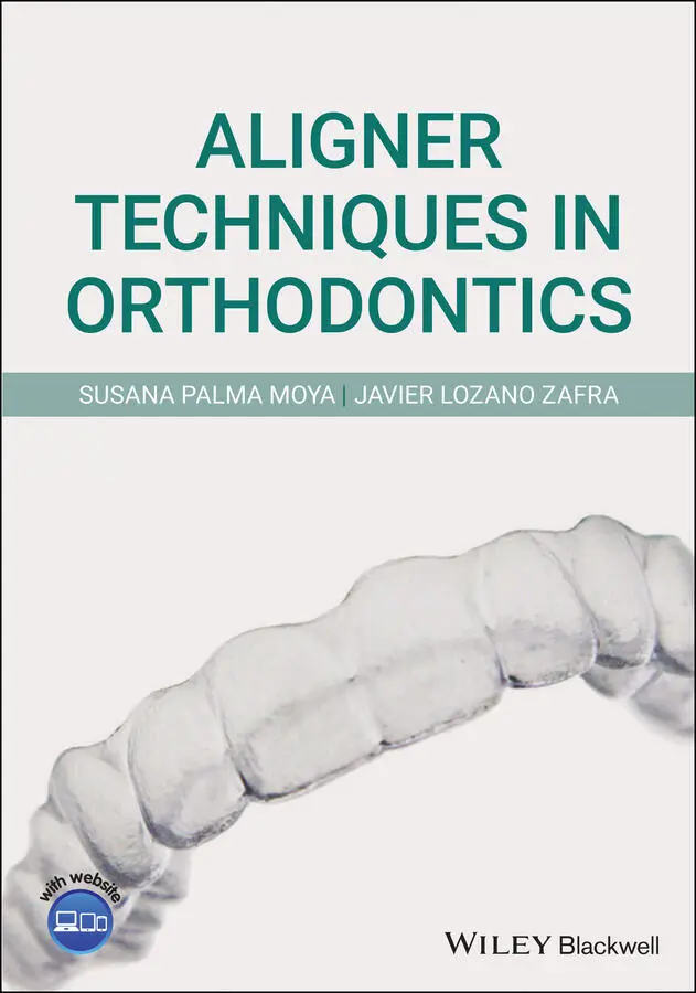
- Название:Aligner Techniques in Orthodontics
- Автор:
- Жанр:
- Год:неизвестен
- ISBN:нет данных
- Рейтинг книги:4 / 5. Голосов: 1
-
Избранное:Добавить в избранное
- Отзывы:
-
Ваша оценка:
Aligner Techniques in Orthodontics: краткое содержание, описание и аннотация
Предлагаем к чтению аннотацию, описание, краткое содержание или предисловие (зависит от того, что написал сам автор книги «Aligner Techniques in Orthodontics»). Если вы не нашли необходимую информацию о книге — напишите в комментариях, мы постараемся отыскать её.
Provides theoretical and practical clinical information on different aligner techniques including Invisalign Offers clear and simple methods to treat patients using different aligner techniques Explains how to use clear aligners to treat a given malocclusion Written by two renowned experts in Align and Invisalign technology Written for practicing orthodontists and general dentists,
provides an invaluable resource for practicing orthodontists.
Aligner Techniques in Orthodontics — читать онлайн ознакомительный отрывок
Ниже представлен текст книги, разбитый по страницам. Система сохранения места последней прочитанной страницы, позволяет с удобством читать онлайн бесплатно книгу «Aligner Techniques in Orthodontics», без необходимости каждый раз заново искать на чём Вы остановились. Поставьте закладку, и сможете в любой момент перейти на страницу, на которой закончили чтение.
Интервал:
Закладка:
15 Chapter 18Fig. 18.1 Skeletal growth pattern has to be carefully evaluated in growing p...Fig. 18.2 Upper temporary spacing is 3 mm, on average, while tooth discrepan...Fig. 18.3 Severe crowding on temporary teeth leads to future extractions or ...Fig. 18.4 Disjunction and protraction should be performed on EC1, while mand...Fig. 18.5 Anterior crossbite management should be addressed early.Fig. 18.6 Skeletal class II can be achieved before than usual if overjet is ...Fig. 18.7 More simple devices can achieve less objectives than aligners. Fig. 18.8 Upper compression with posterior and anterior crowding.Fig. 18.9 Views with the Hyrax before Invisalign First.Fig. 18.10 Panoramic X‐ray before Hyrax.Fig. 18.11 After the Hyrax and before Invisalign First (10‐year‐old patient)...Fig. 18.12 Panoramic X‐ray before Invisalign First.Fig. 18.13 Upper CC superimposition and instructions to CAD designer.Fig. 18.14 Lower CC superimposition and instructions to CAD designer.Fig. 18.15 Right ClinCheck view, initial situation.Fig. 18.16 Left ClinCheck view, initial situation.Fig. 18.17 Attachments can be seen in several areas of the ClinCheck softwar...Fig. 18.18 Pretreatment intraoral views (left, front, right) before Invisali...Fig. 18.19 Intraoral views (left, front, right) with first aligner.Fig. 18.20 Actual situation in additional aligners.Fig. 18.21 Initial and final occlusal (upper and lower).Fig. 18.22 Initial and final smile. Fig. 18.23 Severe crowding impeding proper lateral incisor eruption both in ...Fig. 18.24 Taking advantage of aligner biomechanics by distalizing the secon...Fig. 18.25 Pretreatment intraoral views (right, front, left, upper, lower)....Fig. 18.26 Pretreatment panoramic X‐ray showing current dentition status and...Fig. 18.27 Initial extraoral views.Fig. 18.28 Pretreatment Clinchecks (right, front, left, upper, lower).Fig. 18.29 Refinement: (left) 32 has erupted and does not fit in the aligner...Fig. 18.30 Refinement: panoramic X‐rays show the evolution of the sagittal m...Fig. 18.31 Refinement development: Both available arch space and transverse ...Fig. 18.32 Refinement: 12, 22 have erupted but their clinical crowns are not...Fig. 18.33 Final Intraoral views (right, front, left, upper, lower) showing ...Fig. 18.34 Final and final smile views show an improvement in arch developme...Fig. 18.35 Lateral X‐ray shows incisor proclination as a result of the mecha... Fig. 18.36 Class II growing patient, Lite treatment. Fig. 18.37 Pretreatment views before phase 1 with Hyrax and D‐gainer.Fig. 18.38 Lateral and panoramic X‐rays before phase 1 with Hyrax and D‐gain... Fig. 18.39 Extraoral and intraoral (right, front, left, upper, lower) views ...Fig. 18.40 Panoramic X‐ray, teleradiograph and cephalometry before Invisalig...Fig. 18.41 Upper CC superimposition and instructions to CAD designer.Fig. 18.42 Lower CC superimposition and instructions to CAD designer.Fig. 18.43 Right ClinCheck view, initial situation.Fig. 18.44 Left ClinCheck view, initial situation.Fig. 18.45 Attachments and IPR can be seen in several areas of the ClinCheck...Fig. 18.46 Intraoral views (right, front, left, upper, lower).Fig. 18.47 Intraoral views (right, front, left, upper, lower).Fig. 18.48 Initial and final occlusal views (upper and lower).Fig. 18.49 Pretreatment and final smile and overjet.Fig. 18.50 Initial and final profiles.Fig. 18.51 Final panoramic and lateral X‐rays. Fig. 18.52 Initial intraoral picture.Fig. 18.53 Pretreatment views.Fig. 18.54 Pretreatment panoramic X‐ray, teleradiograph and cephalometry.Fig. 18.55 Upper CC superimposition and instructions to CAD designer.Fig. 18.56 Lower CC superimposition and instructions to CAD designer.Fig. 18.57 Interproximal reduction necessary to upright lower incisors befor...Fig. 18.58 Right ClinCheck view, initial situation.Fig. 18.59 Left ClinCheck view, initial situation.Fig. 18.60 Pretreatment intraoral views (left, front, right).Fig. 18.61 Month 6 of evolution: intraoral views (left, front, right).Fig. 18.62 Refinement: month 15 of evolution; intraoral views (left, front, ...Fig. 18.63 Final intraoral views (right, front, left, upper, lower).Fig. 18.64 Pretreatment and final smile.Fig. 18.65 Final panoramic and lateral X‐rays. Fig. 18.66 Class III growing patient comprehensive treatment, with 23 includ...Fig. 18.67 Intraoral views (right, front, left, upper, lower).Fig. 18.68 Panoramic X‐ray, teleradiograph, cephalometry.Fig. 18.69 Upper CC superimposition and instructions to CAD designer.Fig. 18.70 Lower CC superimposition and instructions to CAD designer.Fig. 18.71 Right ClinCheck view, initial situation.Fig. 18.72 Left ClinCheck view, initial situation.Fig. 18.73 Interproximal reduction in lower arch to create positive overjet....Fig. 18.74 Opening space for 15 (lateral view).Fig. 18.75 Opening space for 15 (occlusal view).Fig. 18.76 Locatelli to open space for 15 and 23.Fig. 18.77 Situation at the end of first set of aligners.Fig. 18.78 (a–f) Transversal development of the arches.Fig. 18.79 Month 18 of evolution, intraoral views.Fig. 18.80 Final views with15 and 23 in place. Fig. 18.81 By labial. Fig. 18.82 By lingual. Fig. 18.83 Class I deep bite Lite treatment.Fig. 18.84 Pretreatment views (right, front, left, upper, lower).Fig. 18.85 Upper CC superimposition and instructions to CAD designer.Fig. 18.86 Lower CC superimposition and instructions to CAD designer.Fig. 18.87 Attachments and IPR can be seen in several areas of the ClinCheck...Fig. 18.88 Left ClinCheck view, initial situation.Fig. 18.89 Right ClinCheck view, initial situation.Fig. 18.90 Pretreatment panoramic X‐ray, teleradiography and cephalometry.Fig. 18.91 Pretreatment (upper) and final (lower) results.Fig. 18.92 Initial and final occlusal (upper and lower).Fig. 18.93 Pretreatment and final smiles and overjets.Fig. 18.94 Final panoramic and lateral X‐rays. Fig. 18.95 Ectopic palatal canine.Fig. 18.96 Initial intraoral views (right, front, left, upper, lower).Fig. 18.97 Initial extraoral views.Fig. 18.98 Initial intraoral views: traction 1, 2, 3.Fig. 18.99 Initial lateral and panoramic X‐rays.Fig. 18.100 Refinement: intraoral views (right, front, left, upper, lower)....Fig. 18.101 ClinChecks: right, front, left, upper, lower.Fig. 18.102 Traction 4 and 5. Initial traction was delivered with constant f...Fig. 18.103 Refinement 2: intraoral views (right, front, left, upper, lower)...Fig. 18.104 Smile at refinement 2.Fig. 18.105 Refinement 2 ClinChecks (right, front, left, upper, lower).Fig. 18.106 Final intraoral views.Fig. 18.107 Final smile view.Fig. 18.108 Current cephalometric measurements.Fig. 18.109 Ortopantomographs (OPG) 0, 1, 2, 3. Evolution of the canine with... Fig. 18.110 Temporary 52.Fig. 18.111 Initial intraoral views (right, front, left, upper, lower).Fig. 18.112 Pretreatment panoramic X‐ray with wisdom teeth present.Fig. 18.113 Final intraoral views (right, front, left, upper, lower).Fig. 18.114 Initial and final panoramic X‐rays. Right class I was achieved e...Fig. 18.115 Final lateral X‐ray and cephalometric analysis.
16 Chapter 19Fig. 19.1 Classic NiTi archwires were only available on standard sizes and s...Fig. 19.2 Expansion can be defined in a precise way, with the decision about...Fig. 19.3 Expansion with aligners can be defined in between Fa point (blue l...Fig. 19.4 Dentoalveolar expansion can be achieved with aligners, as with bra...Fig. 19.5 In this situation, if we ask for the same amount of expansion in b...Fig. 19.6 Check smile In unilateral masticatory side the patient will expose...Fig. 19.7 Asymmetric expansion.Fig. 19.8 Blue arrows indicate proper posterior torque, white ones show arch...Fig. 19.9 Pearl necklace effect.Fig. 19.10 Smile picture. Fig. 19.11 Symmetric compression causing anterior open bite.Fig. 19.12 Pretreatment extraoral and intraoral views.Fig. 19.13 Initial panoramic X‐ray, teleradiograph and cephalometry.Fig. 19.14 Interproximal reduction.Fig. 19.15 Upper superposition.Fig. 19.16 Lower superposition.Fig. 19.17 Initial right ClinCheck view, with CAD designer instructions.Fig. 19.18 Initial left ClinCheck view, with CAD designer instructions.Fig. 19.19 Comparison of initial and final occlusion.Fig. 19.20 Comparison of initial and final occlusal.Fig. 19.21 Comparison of initial and final smile overjets.Fig. 19.22 Comparison of initial and final smiles.Fig. 19.23 Final panoramic X‐ray and teleradiograph. Fig. 19.24 Edge to edge bite with open bite tendency.Fig. 19.25 Pretreatment intraoral views (right, front, left, upper, lower)....Fig. 19.26 Initial extraoral view: short display.Fig. 19.27 Pretreatment panoramic and lateral X‐rays.Fig. 19.28 Pretreatment ClinChecks (right, front, left, upper, lower).Fig. 19.29 Refinement intraoral views (right, front, left, upper, lower).Fig. 19.30 Refinement smile.Fig. 19.31 Refinement ClinChecks (right, front, left, upper, lower).Fig. 19.32 Final intraoral views (right, front, left, upper, lower).Fig. 19.33 Final smile picture and lateral X-ray. Fig. 19.34 Symmetric compression.Fig. 19.35 Pretreatment intraoral views (right, front, left, upper, lower)....Fig. 19.36 Pretreatment panoramic X‐ray, teleradiograph and cephalometry.Fig. 19.37 Upper CC superimposition and instructions to CAD designer.Fig. 19.38 Lower CC superimposition and instructions to CAD designer.Fig. 19.39 Attachments and IPR can be seen in several areas of the ClinCheck...Fig. 19.40 Right ClinCheck view, initial situation.Fig. 19.41 Left ClinCheck view, initial situation.Fig. 19.42 Views before starting refinement.Fig. 19.43 Final views.Fig. 19.44 Comparison of initial and final occlusal (upper and lower).Fig. 19.45 Final expansion reducing buccal corridors.Fig. 19.46 Final Panoramic X‐ray and teleradiography. Fig. 19.47 Symmetric compression with loss of attachment.Fig. 19.48 Pretreatment extraoral and intraoral views.Fig. 19.49 Pretreatment panoramic X‐ray, teleradiograph and cephalometry.Fig. 19.50 Upper CC superimposition and instructions to CAD designer.Fig. 19.51 Lower CC superimposition and instructions to CAD designer.Fig. 19.52 Interproximal reduction is necessary to upright lower incisors.Fig. 19.53 Right and left ClinCheck view, initial situation.Fig. 19.54 Comparison of pretreatment (upper) and final (lower) views.Fig. 19.55 Comparison of initial and final occlusal.Fig. 19.56 Comparison initial of and final smile and overjet. Fig. 19.57 Posterior bilateral crossbite.Fig. 19.58 Initial extraoral views.Fig. 19.59 Initial intraoral views (right, front, left, upper, lower).Fig. 19.60 Pretreatment Clinchecks: right, front, left, upper, lower.Fig. 19.61 Refinement: intraoral views (right, front, left, upper, lower).Fig. 19.62 Refinement ClinChecks: right, front, left, upper, lower.Fig. 19.63 Refinement 2: intraoral views (right, front, left, upper, lower)....Fig. 19.64 Refinement 2 ClinChecks: right, front, left, upper, lower.Fig. 19.65 Final intraoral views (right, front, left, upper, lower).Fig. 19.66 Pretreatment and final smile. Fig. 19.67 Posterior bilateral compression.Fig. 19.68 Initial intraoral views (right, front, left, upper, lower).Fig. 19.69 Initial intraoral views (right, front, left, upper, lower).Fig. 19.70 Pretreatment ClinChecks: right, front, left, upper, lower. Fig. 19.71 Refinement: intraoral views (right, front, left, upper, lower). Fig. 19.72 Refinement ClinChecks: right, front, left, upper, lower.Fig. 19.73 Refinement ClinChecks: right, front, left, upper, lower.Fig. 19.74 Check first column on both charts to see how much expansion was a...Fig. 19.75 Refinement 2: intraoral views (right, front, left, upper, lower)....Fig. 19.76 Final intraoral views: right, front, left, upper, lower.Fig. 19.77 Pretreatment and final smile.Fig. 19.78 Final lateral, cephalometric analysis and panoramic X-ray. Fig. 19.79 patient with severe periodontal disease.Fig. 19.80 Pretreatment views. Note the critical periodontal situation of up...Fig. 19.81 Initial panoramic X-ray, teleradiograph and cephalometry.Fig. 19.82 Problem list.Fig. 19.83 Treatment plan.Fig. 19.84 It is important to relate clinical view to the Clincheck.Fig. 19.85 Upper and lower CC superimposition and instructions to CAD design...Fig. 19.86 Goal of treatment. Open space by asymmetric upper expansion on th...Fig. 19.87 Right ClinCheck view, initial situation.Fig. 19.88 Left ClinCheck view, initial situation.Fig. 19.89 Pontic to open space for a second upper right canine.Fig. 19.90 Views before refinement.Fig. 19.91 Comparison of initial (upper) with final (lower) occlusion. Compo...Fig. 19.92 Comparison of initial and final occlusal.Fig. 19.93 Comparison of initial and final smile and overjet.Fig. 19.94 Comparison of initial (left) and final panoramic x‐rays (right). ...Fig. 19.95 Final teleradiograph. Fig. 19.96 Asymmetric compression.Fig. 19.97 Pretreatment views.Fig. 19.98 Initial panoramic X‐ray, teleradiograph and cephalometry.Fig. 19.99 left‐side deviation of lower jaw.Fig. 19.100 Upper and lower CC superimposition and instructions to CAD desig...Fig. 19.101 Interproximal reduction is necessary to reduce the lower dental ...Fig. 19.102 Initial views before refinement. The posterior open bite on left...Fig. 19.103 Goal of treatment: correct posterior crossbite from 23 to 27.Fig. 19.104 Initial and final design of the asymmetric expansion of the uppe...Fig. 19.105 Comparison of initial and final occlusion.Fig. 19.106 Final occlusion. Both midlines centred. Unilateral posterior cro...Fig. 19.107 Improvement in the smile and in the final torque of upper inciso...Fig. 19.108 Improvement in the profile by clockwise rotation of the mandible... Fig. 19.109 Posterior crossbite with anterior crowding.Fig. 19.110 Careful diagnosis of the crossbite origin will lead to a great r...Fig. 19.111 Cone beam computed tomography: three dimensional radiological ex...Fig. 19.112 Pretreatment intraoral views (right, front, left, upper, lower)....Fig. 19.113 Pretreatment smile and lateral X‐ray.Fig. 19.114 Pretreatment ClinChecks: right, front, left, upper, lower.Fig. 19.115 Refinement: intraoral views (right, front, left, upper, lower)....Fig. 19.116 Smile after first set of aligners.Fig. 19.117 Refinement ClinChecks: right, front, left, upper, lower.Fig. 19.118 Final intraoral views: right, front, left, upper, lower.Fig. 19.119 Pretreatment and final smile: we can observe how posterior expan...Fig. 19.120 Cone beam computed tomography sections. From left (pretreatment)...Fig. 19.121 Posterior crossbite, with anterior crossbite and edge‐to‐edge oc...Fig. 19.122 Smile and upper and lower arch view.Fig. 19.123 Panoramic and lateral X‐rays.Fig. 19.124 Measurements were performed after iTero scanning and OrthoCad so...Fig. 19.125 Skeletal compression has to be approached with a surgical proced...Fig. 19.126 First phase: initially, the patient had a gingivectomy performed...Fig. 19.127 First phase: initially, the patient had a gingivectomy performed...Fig. 19.128 Upper pre‐miniscrew‐assisted rapid palatal expander ( MARPE ): pos...Fig. 19.129 Right, Front, Left Post miniscrew‐assisted rapid palatal expande...Fig. 19.130 Second phase. Measurements were performed after iTero scanning a...Fig. 19.131 Second phase ClinChecks (front, upper, lower).Fig. 19.132 Third phase: intraoral views (front and upper).Fig. 19.133 After 16 aligners space was closed, palatal suture was still ope...Fig. 19.134 Third phase ClinChecks (front, upper, lower).Fig. 19.135 Final intraoral views: front, upper, lower.Fig. 19.136 Pretreatment and final smile.Fig. 19.137 Initial and final cephalometric measurements, with the final pos...Fig. 19.138 Final panoramic X‐ray. Patient smile was widened and skeletal ba...
Читать дальшеИнтервал:
Закладка:
Похожие книги на «Aligner Techniques in Orthodontics»
Представляем Вашему вниманию похожие книги на «Aligner Techniques in Orthodontics» списком для выбора. Мы отобрали схожую по названию и смыслу литературу в надежде предоставить читателям больше вариантов отыскать новые, интересные, ещё непрочитанные произведения.


Обсуждение, отзывы о книге «Aligner Techniques in Orthodontics» и просто собственные мнения читателей. Оставьте ваши комментарии, напишите, что Вы думаете о произведении, его смысле или главных героях. Укажите что конкретно понравилось, а что нет, и почему Вы так считаете.