Pathology of Genetically Engineered and Other Mutant Mice
Здесь есть возможность читать онлайн «Pathology of Genetically Engineered and Other Mutant Mice» — ознакомительный отрывок электронной книги совершенно бесплатно, а после прочтения отрывка купить полную версию. В некоторых случаях можно слушать аудио, скачать через торрент в формате fb2 и присутствует краткое содержание. Жанр: unrecognised, на английском языке. Описание произведения, (предисловие) а так же отзывы посетителей доступны на портале библиотеки ЛибКат.

- Название:Pathology of Genetically Engineered and Other Mutant Mice
- Автор:
- Жанр:
- Год:неизвестен
- ISBN:нет данных
- Рейтинг книги:3 / 5. Голосов: 1
-
Избранное:Добавить в избранное
- Отзывы:
-
Ваша оценка:
Pathology of Genetically Engineered and Other Mutant Mice: краткое содержание, описание и аннотация
Предлагаем к чтению аннотацию, описание, краткое содержание или предисловие (зависит от того, что написал сам автор книги «Pathology of Genetically Engineered and Other Mutant Mice»). Если вы не нашли необходимую информацию о книге — напишите в комментариях, мы постараемся отыскать её.
An updated and comprehensive reference to pathology in every organ system in genetically modified mice Pathology of Genetically Engineered and Other Mutant Mice
Pathology of Genetically Engineered and Other Mutant Mice
Pathology of Genetically Engineered and Other Mutant Mice — читать онлайн ознакомительный отрывок
Ниже представлен текст книги, разбитый по страницам. Система сохранения места последней прочитанной страницы, позволяет с удобством читать онлайн бесплатно книгу «Pathology of Genetically Engineered and Other Mutant Mice», без необходимости каждый раз заново искать на чём Вы остановились. Поставьте закладку, и сможете в любой момент перейти на страницу, на которой закончили чтение.
Интервал:
Закладка:
The sensory functions of primary cilia depend on the coordinated trafficking and temporal localization of specific receptors and their associated signal transduction modules in the cilium. Primary cilia sense and transduce environmental signals via several signaling pathways, including HH, GPCR, WNT, receptor tyrosine kinase, and transforming growth factor β (TGFβ)/bone morphogenetic protein (BMP) signaling [8]. The central importance of these pathways in regulating key processes in development and tissue homeostasis account for the pleomorphic developmental disorders and diseases that accompany primary cilia defects [45]. The role of primary cilia in HH signaling is probably the best understood at this time. There are three major protein ligands in the HH system: SHH, Indian Hedgehog (IHH), and Desert Hedgehog (DHH). Of these, SHH signaling is critical to spatial patterning of the neuroepithelium, cellular identity in the CNS, axonal guidance, wiring of the neural network, and neuronal activity. Primary cilia defects in mice with mutations in SHH are associated with several defects in brain development, such as defective neural patterning, cerebellar hypoplasia, and defective hippocampal neurogenesis. IHH and DHH are both essential for normal craniofacial development, and mutations in several HH‐related ciliary proteins result in skeletal and craniofacial deformities [46].
Cystic Kidney Diseases
PKDs were the first to be linked definitively to defects in primary immotile cilia. Recognition that polycystin 1 (PKD1), polycystin 2 (PKD2), and nephrocystin 1 (NPHP1) proteins were all localized to the primary cilium/basal body/centrosome provided the first evidence that dysfunctional primary cilia could be involved in the pathogenesis of PKD and nephronophthisis (NPHP) [47, 48]. Since then mutations in dozens of other cilia‐related genes have been directly linked to the development of cystic kidney diseases [49]. The primary cilium on renal tubular epithelial cells projects into the urinary space and appears to act as a mechanosensor in detecting the flow of urine and thereby influencing renal cell division [50]. The primary cilium also regulates the orientation of cell division and disrupted planar cell polarity signaling also contributes to renal cystogenesis [51]. PKD is characterized by grossly enlarged kidneys, due to markedly dilated renal tubules lined by epithelial cells showing increased mitotic activity ( Figure 6.4). In contrast, the polycystic kidneys in NPHP are essentially normal or reduced in size, with low mitotic rates and increased apoptosis of renal tubular epithelium [52] ( Figure 6.5).
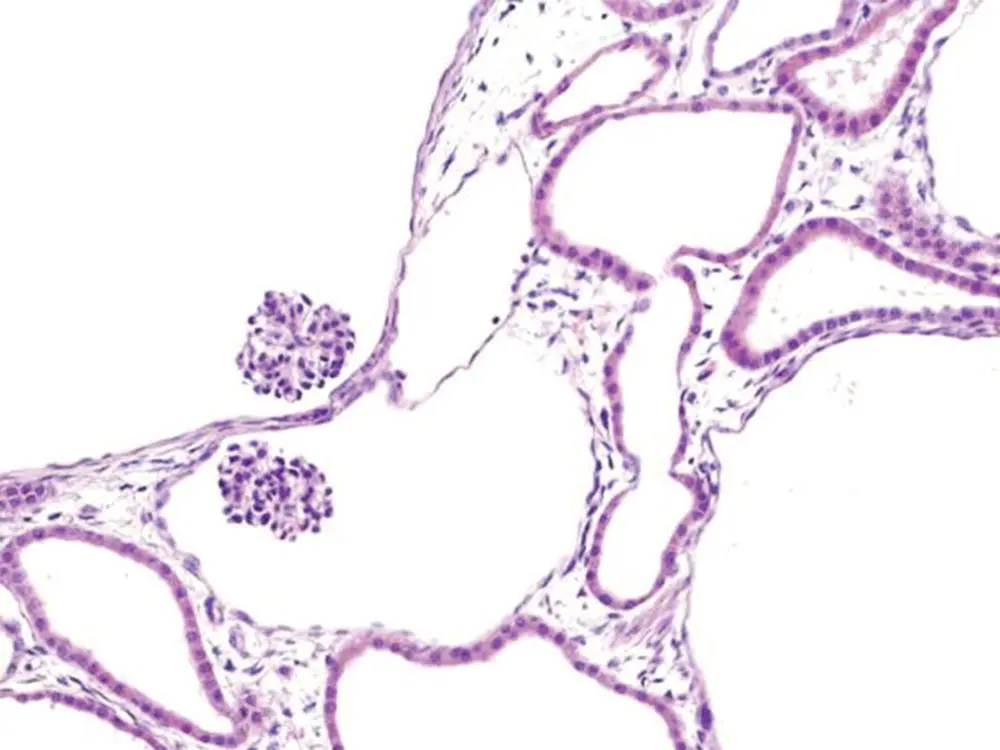
Figure 6.4 Polycystic kidney disease. Dilatation of renal tubules in an enlarged kidney due to proliferation of renal epithelium is associated with dysfunctional primary cilia.
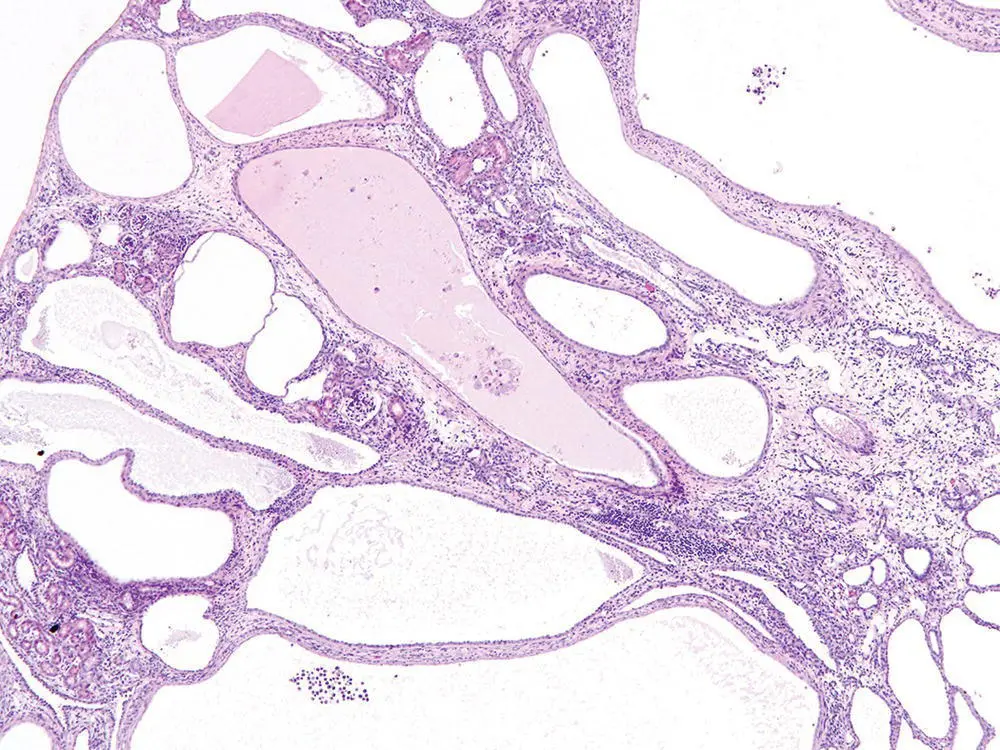
Figure 6.5 Nephronophthisis. Cystic tubules with interstitial inflammation and fibrosis in a normal sized kidney are associated with attrition of renal epithelium due to dysfunctional primary cilia.
Retinal Degeneration
Retinal degeneration is the most common manifestation of ciliary dysfunction in the eye, although coloboma and microphthalmia can occur in some ciliopathies as a result of defective HH signaling. In the retinal photoreceptor cells, modified primary cilia form the photoreceptor outer segments and express proteins that are specialized for phototransduction. The outer segments are specialized sensory cilia, and deficient morphogenesis and/or dysfunction of these sensory cilia caused by mutations in a wide range of photoreceptor‐specific and ciliary genes can result in inherited retinal degenerations [53]. Almost all of the proteins involved in maintenance of the outersegment and phototransduction (most notably rhodopsin) are synthesized in the inner segment and must be transported to the outer segment via the connecting cilium. As a result, the efficient transport of cargo along the connecting cilium is essential for the assembly and maintenance of the photoreceptor outer segment. Mutations in several genes that encode proteins localized to the connecting cilium and/or its basal body can disrupt this process of intersegmental transport and result in the mislocalization of outer segment proteins and disorganization of the outer segments [54–58], which often precedes photoreceptor degeneration [59, 60]. In addition, the axoneme of the connecting cilium extends into the OS and is thought to establish the proper alignment of the membranous disks containing rhodopsin and other proteins required for phototransduction [61].
Histology is a very sensitive and specific method for detecting retinopathies. In normal mouse eyes, subretinal macrophages are absent or very rare, the inner and outer segments of the photoreceptors appear in organized columns and are of approximately equal length, and the outer nuclear layer is thicker than the inner nuclear layer. Almost all disruptions affecting the development or function of outer segments will lead to degeneration of photoreceptors, which can be detected histologically even at early stages by the appearance or any combination of the following changes: increased numbers of subretinal macrophages ( Figure 6.6), disrupted/thin outer segments, or reduced thickness of the outer nuclear layer (consisting of photoreceptor nuclei; Figure 6.7).
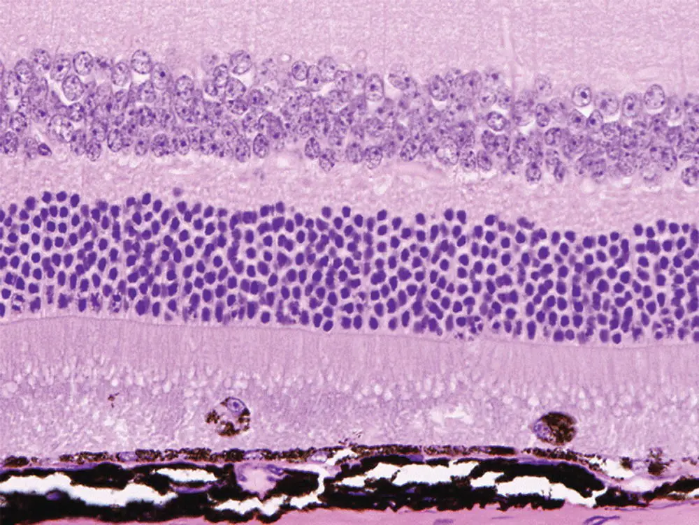
Figure 6.6 Eye. Macrophages within the outer segment of photoreceptor are present at the earlier stages of retinal degeneration.
Heart Defects
Careful gross examination of the heart and great vessels should be included when phenotyping any genetically engineered mouse line showing decreased viability [62, 63]. Cilia play a key role in cell signaling pathways that regulate the development of the heart and large blood vessels, particularly those involved in left–right patterning. Although situs inversus totalis is generally asymptomatic, complex congenital cardiac defects and transposition of the great vessels of the heart are often associated with situs ambiguus or heterotaxy [64–66]. Interestingly, humans with congenital heart disease show a high prevalence of ciliary dysfunction and de novo ciliary mutations, suggesting that ciliary functions other than left–right positioning may be involved [67].
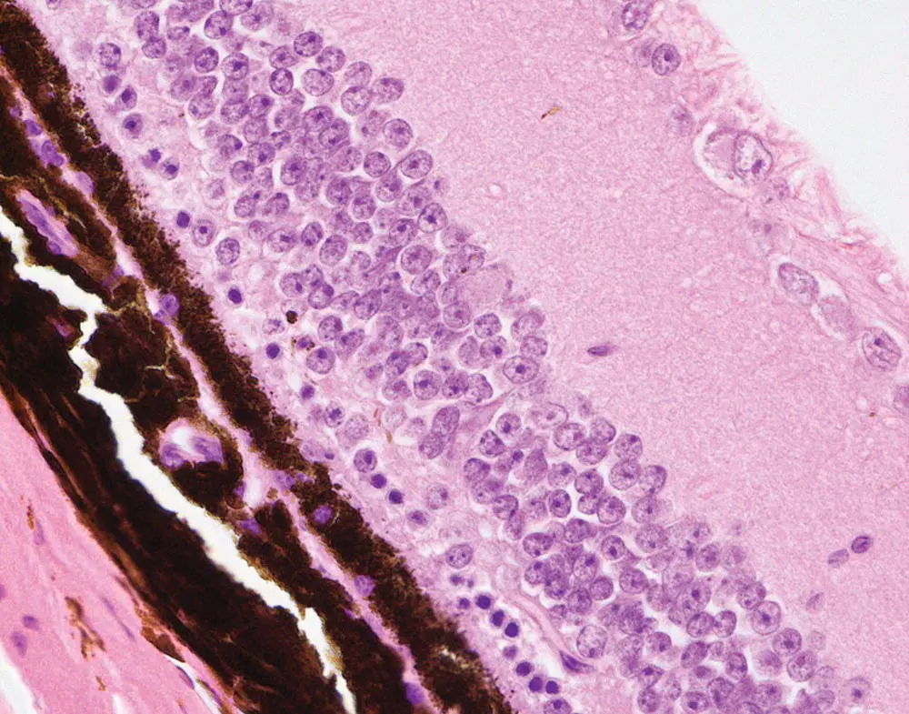
Figure 6.7 Eye. Advanced retinal degeneration there is characterized by a marked reduction in the thickness of the outer nuclear layer photoreceptors.
Skeletal Defects
Ciliopathies affecting skeletal development most often appear as malformed ribs, polydactyly, brachydactyly, shortened long bones, and cone‐shaped epiphyses. Disruption of HH signaling regulation leads to multiple bone diseases [68] and many of the mutated genes associated with these skeletal defects encode proteins involved ciliary transport and thus HH signaling [69, 70]. SHH is a major morphogen during early stages of embryonic limb development, while IHH has a key role in endochondral ossification.
Читать дальшеИнтервал:
Закладка:
Похожие книги на «Pathology of Genetically Engineered and Other Mutant Mice»
Представляем Вашему вниманию похожие книги на «Pathology of Genetically Engineered and Other Mutant Mice» списком для выбора. Мы отобрали схожую по названию и смыслу литературу в надежде предоставить читателям больше вариантов отыскать новые, интересные, ещё непрочитанные произведения.


Обсуждение, отзывы о книге «Pathology of Genetically Engineered and Other Mutant Mice» и просто собственные мнения читателей. Оставьте ваши комментарии, напишите, что Вы думаете о произведении, его смысле или главных героях. Укажите что конкретно понравилось, а что нет, и почему Вы так считаете.