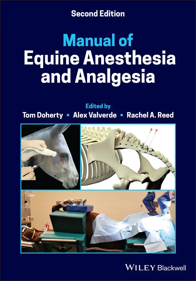
Manual of Equine Anesthesia and Analgesia
Здесь есть возможность читать онлайн «Manual of Equine Anesthesia and Analgesia» — ознакомительный отрывок электронной книги совершенно бесплатно, а после прочтения отрывка купить полную версию. В некоторых случаях можно слушать аудио, скачать через торрент в формате fb2 и присутствует краткое содержание. Жанр: unrecognised, на английском языке. Описание произведения, (предисловие) а так же отзывы посетителей доступны на портале библиотеки ЛибКат.
- Название:Manual of Equine Anesthesia and Analgesia
- Автор:
- Жанр:
- Год:неизвестен
- ISBN:нет данных
- Рейтинг книги:3 / 5. Голосов: 1
-
Избранное:Добавить в избранное
- Отзывы:
-
Ваша оценка:
Manual of Equine Anesthesia and Analgesia: краткое содержание, описание и аннотация
Предлагаем к чтению аннотацию, описание, краткое содержание или предисловие (зависит от того, что написал сам автор книги «Manual of Equine Anesthesia and Analgesia»). Если вы не нашли необходимую информацию о книге — напишите в комментариях, мы постараемся отыскать её.
Provides practical, clinically oriented information on anesthetizing equids Uses a bulleted format designed for fast access of key information Offers step-by-step instructions and diagrams of nerve blocks of the limbs, head, and ophthalmic structures Includes new coverage of topics including regulation of extracellular fluid and blood pressure, acid-base disorders, and hemodynamic effects of autonomic drugs
remains a must-have resource for all equine practitioners and veterinary students involved with anesthetizing horses.
Manual of Equine Anesthesia and Analgesia — читать онлайн ознакомительный отрывок
Ниже представлен текст книги, разбитый по страницам. Система сохранения места последней прочитанной страницы, позволяет с удобством читать онлайн бесплатно книгу «Manual of Equine Anesthesia and Analgesia», без необходимости каждый раз заново искать на чём Вы остановились. Поставьте закладку, и сможете в любой момент перейти на страницу, на которой закончили чтение.
Интервал:
Закладка:
5 Brosnan, R.J., Steffey, E.P., LeCouteur, R.A. et al. (2002). Effects of body position on intracranial and cerebral perfusion pressures in isoflurane‐anesthetized horses. J. Appl. Physiol. 92: 2542–2546.
6 Brosnan, R.J., Esteller‐Vico, A., Steffey, E.P. et al. (2008). Effects of head‐down positioning on regional central nervous system perfusion in isoflurane‐anesthetized horses. Am. J. Vet. Res. 69: 737–743.
7 Brosnan, R.J., Steffey, E.P., LeCouteur, R.A. et al. (2011). Effects of isoflurane anesthesia on cerebrovascular autoregulation in horses. Am. J. Vet. Res. 72: 18–24.
8 Caines, D., Sinclair, M., Valverde, A. et al. (2014). Comparison of isoflurane and propofol for maintenance of anesthesia in dogs with intracranial disease undergoing magnetic resonance imaging. Vet. Anaesth. Analg. 41: 468–479.
9 Chaffin, M.K., Walker, M.A., McArthur, N.H. et al. (1997). Magnetic resonance imaging of the brain of normal neonatal foals. Vet. Radiol. Ultrasound 38: 102–111.
10 Drynan, E.A., Gray, P., and Raisis, A. (2012). Incidence of seizures associated with the use of acepromazine in dogs undergoing myelography. J. Vet. Crit. Care 22: 262–266.
11 Duke‐Novakovski, T., Palacios‐Jiminez, C., Wetzel, T. et al. (2015). Cardiopulmonary effects of dexmedetomidine and ketamine infusions with either propofol infusion or isoflurane for anesthesia in horses. Vet. Anaesth. Analg. 42: 39–49.
12 Farrell, D. and Bendo, A.A. (2018). Perioperative management of severe traumatic brain injury: What Is New? Curr. Anesthesiol. Rep. 8: 279–289.
13 Flaherty, D., Reid, J., Welsh, E. et al. (1997). A pharmacodynamic study of propofol or propofol and ketamine infusions in ponies undergoing surgery. Res. Vet. Sci. 62: 179–184.
14 Hirota, K., Hashimoto, Y., Sato, T. et al. (2001). Bronchoconstrictive and relaxant effects of lidocaine on the airway in dogs. Crit. Care Med. 29: 1040–1044.
15 Jones, T., Bracamonte, J.L., Ambros, B., and Duke‐Novakovski, T. (2019). Total intravenous anesthesia with alfaxalone, dexmedetomidine and remifentanil in healthy foals undergoing abdominal surgery. Vet. Anaesth. Analg. 46: 315–324.
16 Kortz, G.D., Madigan, J.E., Goetzman, B.W., and Durando, M. (1995). Intracranial pressure and cerebral perfusion in clinically normal equine neonates. Am. J. Vet. Res. 56: 1351–1355.
17 Lacombe, V.A. (2015). Seizures in horses: diagnosis and classification. Vet. Med. Res. Reports 6: 301–308.
18 McConnell, J., Kirby, R., and Rudloff, E. (2007). Administration of acepromazine maleate to 31 dogs with a history of seizures. J. Vet. Emerg. Crit. Care 17: 262–267.
19 Miyazaki, Y., Adachi, T., Kurata, J. et al. (1999). Dexmedetomidine reduces seizure threshold during enflurane anaesthesia in cats. Br. J. Anaesth. 82: 935–937.
20 Rasmussen, N.J., Rosendal, T., and Overgaard, J. (1978). Althesin in neurosurgical patients: effects on cerebral hemodynamics and metabolism. Acta Anaesthesiol. Scand. 22: 257–269.
21 Sande, A. and West, C. (2010). Traumatic brain injury: a review of pathophysiology and management. J. Vet. Emerg. Crit. Care 20: 177–190.
22 Serrano, S., Hughes, D., and Chandler, K. (2006). Use of ketamine for the management of refractory status epilepticus in a dog. J. Vet. Intern. Med. 20: 194–197.
23 Spadavecchia, C., Jaggy, A., Fatzer, R., and Schatzmann, U. (2001). Postanaesthetic cerebral necrosis in a horse. Equine Vet. J. 33: 621–624.
24 Steffen, F. and Grasmueck, S. (2000). Propofol for treatment of refractory seizures in dogs and a cat with intracranial disorders. J. Small Anim. Pract. 41: 496–499.
25 Talke, P., Tong, C., Lee, H.W. et al. (1997). Effect of dexmedetomidine on lumbar cerebrospinal fluid pressure in humans. Anesth. Analg. 85: 358–364.
26 Tobias, K.M. and Marioni‐Henry, K.A. (2006). Retrospective study on the use of acepromazine maleate in dogs with seizures. J. Am. Anim. Hosp. Assoc. 42: 283–289.
27 Warne, L.N., Beths, T., Fogal, S., and Bauquier, S.H. (2014). The use of alfaxalone and remifentanil total intravenous anesthesia in a dog undergoing a craniectomy for tumor resection. Can. Vet. J. 55: 1083–1088.
28 Zornow, M.H., Fleischer, J.E., Sceller, M.S. et al. (1990). Dexmedetomidine, an alpha 2‐adrenergic agonist, decreases cerebral blood flow in the isoflurane‐anesthetized dog. Anesth. Analg. 70: 624–630.
7 The Autonomic Nervous System
Christine Egger
The autonomic nervous system (ANS) is the portion of the central nervous system (CNS) that controls visceral functions such as arterial blood pressure, heart rate, respiratory function, gastrointestinal (GI) motility and secretion, urinary bladder emptying, sweating, body temperature, as well as a number of other important bodily functions.
I General organization of the ANS (functional anatomy)
The ANS is anatomically divided into the central and peripheral nervous system, and functionally divided into the sympathetic (adrenergic) nervous system (SNS) and the parasympathetic (cholinergic) nervous system (PNS).
A Central autonomic nervous system
The hypothalamus is the principle site of ANS integration (e.g. blood pressure control, thermoregulation, stress response).
The medulla oblongata and pons contain the centers for hemodynamic and ventilatory control.
B Peripheral autonomic nervous system
Pre‐ganglionic neurons are myelinated, rapidly conducting, originate in the CNS, and synapse in an autonomic ganglion.
Post‐ganglionic neurons are unmyelinated, slower conducting, arise from the autonomic ganglia, and are distributed to effector organs.
The SNS (paravertebral) ganglia are located nearer to the spinal cord than to the innervated organ, and the PNS ganglia are located in or near the innervated organ.
C Afferent input
The ANS centers in the brain stem act as relay stations for control of activities initiated at higher levels of the brain, such as the hypothalamus and cerebrum.
Visceral sensory signals entering the autonomic ganglia, spinal cord, brain stem, or hypothalamus elicit reflex responses which control the activity of visceral organs.
D Efferent output
Efferent autonomic signals are transmitted to the body through two major subdivisions of the ANS, the SNS, and PNS. (see later discussion.)
II Physiology of the autonomic nervous system
A Neurotransmitters
Acetylcholine (ACh) and norepinephrine (NE) are the main neurotransmitters.
All preganglionic neurons are cholinergic in both the SNS and PNS nervous systems and secrete ACh.
Postganglionic neurons of the PNS are cholinergic, while most postganglionic SNS neurons are adrenergic and secrete NE.The adrenal gland functions like a modified sympathetic ganglion and when stimulated by presynaptic SNS fibers, releases NE and epinephrine (EPI) into the circulation to act on sites distant from the gland; therefore, there is no postganglionic neuron.Sweat glands in horses are mostly apocrine and open into hair follicles. These glands and the piloerector muscles are stimulated by circulating EPI that acts on adrenergic (β2) receptors.Eccrine sweat glands are common in humans, but are less common in horses. In humans, they constitute 90% of sweat glands and they are stimulated by a postganglionic SNS neuron that secretes ACh.
B Synthesis, duration of action, and degradation of ACh
ACh is synthesized within the axoplasm in the terminal endings of cholinergic nerves.
The ACh is transported to the interior of the vesicles where it is stored in a highly concentrated form until it is released along with ATP.
Читать дальшеИнтервал:
Закладка:
Похожие книги на «Manual of Equine Anesthesia and Analgesia»
Представляем Вашему вниманию похожие книги на «Manual of Equine Anesthesia and Analgesia» списком для выбора. Мы отобрали схожую по названию и смыслу литературу в надежде предоставить читателям больше вариантов отыскать новые, интересные, ещё непрочитанные произведения.


Обсуждение, отзывы о книге «Manual of Equine Anesthesia and Analgesia» и просто собственные мнения читателей. Оставьте ваши комментарии, напишите, что Вы думаете о произведении, его смысле или главных героях. Укажите что конкретно понравилось, а что нет, и почему Вы так считаете.