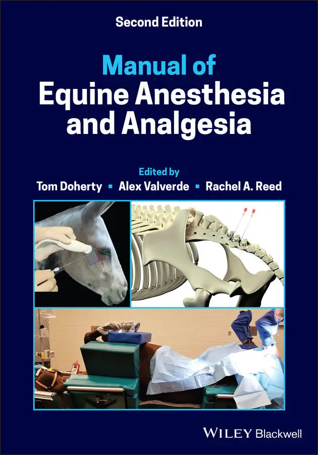
Manual of Equine Anesthesia and Analgesia
Здесь есть возможность читать онлайн «Manual of Equine Anesthesia and Analgesia» — ознакомительный отрывок электронной книги совершенно бесплатно, а после прочтения отрывка купить полную версию. В некоторых случаях можно слушать аудио, скачать через торрент в формате fb2 и присутствует краткое содержание. Жанр: unrecognised, на английском языке. Описание произведения, (предисловие) а так же отзывы посетителей доступны на портале библиотеки ЛибКат.
- Название:Manual of Equine Anesthesia and Analgesia
- Автор:
- Жанр:
- Год:неизвестен
- ISBN:нет данных
- Рейтинг книги:3 / 5. Голосов: 1
-
Избранное:Добавить в избранное
- Отзывы:
-
Ваша оценка:
Manual of Equine Anesthesia and Analgesia: краткое содержание, описание и аннотация
Предлагаем к чтению аннотацию, описание, краткое содержание или предисловие (зависит от того, что написал сам автор книги «Manual of Equine Anesthesia and Analgesia»). Если вы не нашли необходимую информацию о книге — напишите в комментариях, мы постараемся отыскать её.
Provides practical, clinically oriented information on anesthetizing equids Uses a bulleted format designed for fast access of key information Offers step-by-step instructions and diagrams of nerve blocks of the limbs, head, and ophthalmic structures Includes new coverage of topics including regulation of extracellular fluid and blood pressure, acid-base disorders, and hemodynamic effects of autonomic drugs
remains a must-have resource for all equine practitioners and veterinary students involved with anesthetizing horses.
Manual of Equine Anesthesia and Analgesia — читать онлайн ознакомительный отрывок
Ниже представлен текст книги, разбитый по страницам. Система сохранения места последней прочитанной страницы, позволяет с удобством читать онлайн бесплатно книгу «Manual of Equine Anesthesia and Analgesia», без необходимости каждый раз заново искать на чём Вы остановились. Поставьте закладку, и сможете в любой момент перейти на страницу, на которой закончили чтение.
Интервал:
Закладка:
17 Chapter 23Figure 23.1 Anatomy of the major motor and sensory nerves of the equine peri...Figure 23.2 Sites of equine periocular nerve blocks. 1: auriculopalpebral; 2...Figure 23.3 Approximate areas of desensitization afforded by periocular sens...Figure 23.4 Locating the equine supraorbital foramen using Töth's law.Figure 23.5 Placement of spinal needle for supraorbital fossa block.
18 Chapter 24Figure 24.1 Subcircumneural space surrounding the nerve, artery, and vein wi...Figure 24.2 The neurovascular bundle containing the palmar/plantar digital n...Figure 24.3 The neurovascular bundle containing the palmar/plantar digital n...Figure 24.4 Location of needle insertion for the low four‐point nerve block....Figure 24.5 Desensitization of palmar/plantar nerves distal to the ramus com ...Figure 24.6 Location of needle insertion for four‐point (high palmar) nerve ...Figure 24.7 Location of needle insertion for the lateral palmar nerve block ...Figure 24.8 Location of needle insertion for blockade of the median nerve an...Figure 24.9 Location of needle insertion for blockade of the ulnar nerve pro...Figure 24.10 Location of needle insertion for the high plantar nerve block....Figure 24.11 Location of needle insertion for the deep branch of the lateral...Figure 24.12 Location of needle insertion for blockade of the tibial nerve....Figure 24.13 Location of needle insertion for blockade of the peroneal nerve...
19 Chapter 25Figure 25.1 Dissection of the pelvic cavity showing the pudendal nerve stain...Figure 25.2 Caudal aspect of the pelvic cavity displaying the superficial pe...Figure 25.3 Landmarks for the pudendal nerve block in the horse. The white d...Figure 25.4 Peripheral nerve locator needle positioning in a mare.Figure 25.5 Intratesticular injection of 2% mepivacaine (Carbocaine®).
20 Chapter 26Figure 26.1 First step, localizing the transverse process of L3.Figure 26.2 Left thoracolumbar area of a standing adult Thoroughbred horse f...Figure 26.3 Red stars indicate location of nerve roots of T18, L1, and L2. N...Figure 26.4 Anatomic location of spinal nerve roots T18, L1, L2, and L3 and ...Figure 26.5 Use of caudal border of last rib to determine location of third ...Figure 26.6 Insertion of needle for blind paravertebral block.Figure 26.7 Aspirate prior to injection to ensure that the needle tip is not...Figure 26.8 Transversus abdominus plane block using the flank approach.Figure 26.9 Transversus abdominus plane block using the intercostal ventral ...Figure 26.10 Transversus abdominus plane block using the subcostal approach....Figure 26.11 Caudal intercostal block performed in the standing horse for ab...
21 Chapter 27Figure 27.1 Palpation of the sacrococcygeal (S‐Co) and intercoccygeal space ...Figure 27.2 Superficial infiltration of local anesthetic using a 23–25‐gauge...Figure 27.3 (epidural with spinal cord included ). A caudal epidural using th...Figure 27.4 (epidural without spinal cord included) . A caudal epidural using...Figure 27.5 (epidural catheter) . An epidural catheter placed at the first in...
22 Chapter 29Figure 29.1 Pain scoring record on stall door of equine patient in hospital....Figure 29.2 The top row displays pictures of horses that are not in pain. Th...Figure 29.3 An example of low‐level intensity “rolling.” This horse later pr...Figure 29.4 An example of the gross pain behavior “mouth playing.”Figure 29.5 This horse is not in a normal resting position as the front limb...Figure 29.6 An attentive horse standing in the front of the box stall – norm...Figure 29.7 A horse, standing in the front of the box stall with no attentio...Figure 29.8 This horse is “tucked up,” there is tension of the abdominal wal...Figure 29.9 This horse is not weight bearing on the left front limb. Notice ...Figure 29.10 This horse had wound surgery on the right hind limb not many ho...Figure 29.11 This horse shows attention toward the painful area. In this cas...Figure 29.12 This horse was lame. The stifle was the reason for the lameness...Figure 29.13 This horse had wound surgery in the hind limb but also very bri...Figure 29.14 Horses in severe pain or long‐term pain, may not show any inter...
23 Chapter 30Figure 30.1 Goniometer assessing the range of motion of the left carpus.Figure 30.2 Pressure algometer assessing the mechanical nociceptive threshol...Figure 30.3 Class 4 therapeutic laser.Figure 30.4 Electrical stimulation of the right gluteal.Figure 30.5 Extracorporeal shockwave therapy of the right proximal metacarpa...Figure 30.6 A selection of acupuncture needles.Figure 30.7 Electroacupuncture and dry needle treatment for support limb lam...Figure 30.8 Electroacupuncture for cervical pain.Figure 30.9 Electroacupuncture for thoracolumbar and pelvic area pain.Figure 30.10 Electroacupuncture treatment for sacrococcygeal injury with uri...
24 Chapter 31Figure 31.1 Foal anesthetized and breathing spontaneously immediately post i...Figure 31.2 Positioning and padding to support limbs of a foal and protect b...Figure 31.3 Hand recovering a foal. One person supports the head and another...Figure 31.4 Mare and foal in induction area. The foal will be induced with t...Figure 31.5 Repair of a tear in urinary bladder.Figure 31.6 Urine in suction jar after removal from abdomen. It is important...
25 Chapter 32Figure 32.1 Distended and fluid‐filled small intestine with compromised bloo...Figure 32.2 Use of a demand valve to ventilate a horse after induction.Figure 32.3 Horse on pads in recovery. An orotracheal tube is in place.
26 Chapter 33Figure 33.1 22‐year‐old horse with Cushing's disease ( pituitary pars interme ...
27 Chapter 34Figure 34.1 Delivery of live foal, lifting by hindlimbs out of uterus.Figure 34.2 Delivery of live foal; hindlimbs, abdomen, and thorax exiting ut...Figure 34.3 Positioning of mare for attempted vaginal delivery of the fetus....
28 Chapter 35Figure 35.1 Warning signage posted outside of the MRI room.Figure 35.2 Custom cutout of the gantry of a 3.0 Tesla MRI allows for the ho...Figure 35.3 Low‐field MRI used for imaging of standing horses.Figure 35.4 Table used for equine CT under general anesthesia.Figure 35.5 Stocks used for CT in the standing horse with open side.Figure 35.6 Sandbags used to limit motion of head and neck during image acqu...Figure 35.7 Platform for horse to stand on during standing CT image acquisit...Figure 35.8 Horse's head in CT gantry.
29 Chapter 36Figure 36.1 Pharyngeal/laryngeal anatomy of the donkey;(a) arytenoid cartila...Figure 36.2 Jugular vein traveling deep to the cutaneous colli muscle.
30 Chapter 37Figure 37.1 Use of a Dan‐inject rifle to dart a wild horse.Figure 37.2 Delivery of supplemental inspired oxygen in the field. An E‐cyli...Figure 37.3 Wild horse restrained in a chute system to facilitate intramuscu...
31 Chapter 38Figure 38.1 Multiparameter anesthetic monitor indicating pulse pressure vari...Figure 38.2 Horse with radial nerve injury following recovery from anesthesi...Figure 38.3 Horse with femoral nerve injury following recovery from anesthes...Figure 38.4 Horse with facial nerve injury following recovery from anesthesi...Figure 38.5 Priapism in standing horse following recovery from general anest...Figure 38.6 Swelling of the distal portion of the penis secondary to constri...Figure 38.7 Fibrosis of the penis subsequent to trauma and infection followi...Figure 38.8 Caudal curvature of the penis as a consequence of fibrosis.Figure 38.9 Bandaging of the penis to the abdomen to reduce edema formation....Figure 38.10 Probang ready for insertion into prepuce.Figure 38.11 Probang in position to retain the penis in the preputial cavity...Figure 38.12 Image of retainer bottle to be used as a probang.Figure 38.13 Image of retainer bottle in position in prepuce.Figure 38.14 Urticarial lesion on a horse. The horse was sedated with xylazi...
Читать дальшеИнтервал:
Закладка:
Похожие книги на «Manual of Equine Anesthesia and Analgesia»
Представляем Вашему вниманию похожие книги на «Manual of Equine Anesthesia and Analgesia» списком для выбора. Мы отобрали схожую по названию и смыслу литературу в надежде предоставить читателям больше вариантов отыскать новые, интересные, ещё непрочитанные произведения.


Обсуждение, отзывы о книге «Manual of Equine Anesthesia and Analgesia» и просто собственные мнения читателей. Оставьте ваши комментарии, напишите, что Вы думаете о произведении, его смысле или главных героях. Укажите что конкретно понравилось, а что нет, и почему Вы так считаете.