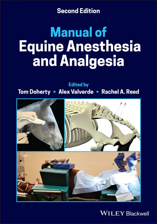
Manual of Equine Anesthesia and Analgesia
Здесь есть возможность читать онлайн «Manual of Equine Anesthesia and Analgesia» — ознакомительный отрывок электронной книги совершенно бесплатно, а после прочтения отрывка купить полную версию. В некоторых случаях можно слушать аудио, скачать через торрент в формате fb2 и присутствует краткое содержание. Жанр: unrecognised, на английском языке. Описание произведения, (предисловие) а так же отзывы посетителей доступны на портале библиотеки ЛибКат.
- Название:Manual of Equine Anesthesia and Analgesia
- Автор:
- Жанр:
- Год:неизвестен
- ISBN:нет данных
- Рейтинг книги:3 / 5. Голосов: 1
-
Избранное:Добавить в избранное
- Отзывы:
-
Ваша оценка:
Manual of Equine Anesthesia and Analgesia: краткое содержание, описание и аннотация
Предлагаем к чтению аннотацию, описание, краткое содержание или предисловие (зависит от того, что написал сам автор книги «Manual of Equine Anesthesia and Analgesia»). Если вы не нашли необходимую информацию о книге — напишите в комментариях, мы постараемся отыскать её.
Provides practical, clinically oriented information on anesthetizing equids Uses a bulleted format designed for fast access of key information Offers step-by-step instructions and diagrams of nerve blocks of the limbs, head, and ophthalmic structures Includes new coverage of topics including regulation of extracellular fluid and blood pressure, acid-base disorders, and hemodynamic effects of autonomic drugs
remains a must-have resource for all equine practitioners and veterinary students involved with anesthetizing horses.
Manual of Equine Anesthesia and Analgesia — читать онлайн ознакомительный отрывок
Ниже представлен текст книги, разбитый по страницам. Система сохранения места последней прочитанной страницы, позволяет с удобством читать онлайн бесплатно книгу «Manual of Equine Anesthesia and Analgesia», без необходимости каждый раз заново искать на чём Вы остановились. Поставьте закладку, и сможете в любой момент перейти на страницу, на которой закончили чтение.
Интервал:
Закладка:
19 Chapter 38Table 38.1 Drugs used in management of anaphylactic and anaphylactoid reacti...
20 Chapter 39Table 39.1 Drugs commonly used for sedation and analgesia pre‐recovery and d...Table 39.2 Common techniques of recovery in the horse.Table 39.3 Criteria for selecting method of assisted recovery.
List of Illustrations
1 Chapter 1 Figure 1.1 Rinsing of the mouth with water prior to induction of anesthesia ...
2 Chapter 2 Figure 2.1 Bench top point‐of‐care blood analyzer, Figure 2.2 Stall‐side point‐of‐care blood analyzer,
3 Chapter 3 Figure 3.1 The Wigger's diagram depicts the electrical, mechanical and audib... Figure 3.2 Normal mucous membrane color, blanching of mucous membranes with ... Figure 3.3 Normal sinus rhythm. Figure 3.4 Second‐degree atrioventricular blockade. Figure 3.5 Atrial fibrillation. Figure 3.6 Premature ventricular complex. Figure 3.7 Ventricular bigeminy; every other complex is ventricular in origi... Figure 3.8 Ventricular tachycardia.
4 Chapter 4 Figure 4.1 Normal equine larynx: arytenoid cartilage (a), vocal fold (b), ep... Figure 4.2 Lung volumes in an adult horse (500 kg). TLC, Total lung capacity Figure 4.3 Pulmonary pressure‐volume curve; illustrating greater pressure di... Figure 4.4 Blood entering the pulmonary capillaries associated with non‐vent... Figure 4.5 The oxy‐hemoglobin dissociation curve illustrates the relationshi... Figure 4.6 PVC mouth gag used to hold mouth open and protect endotracheal tu... Figure 4.7 Oral speculums used in equine patients: Weingart mouth gag (a), G... Figure 4.8 Intubation of a horse in sternal recumbency if there is a risk of... Figure 4.9 Horses with orotracheal (a) and nasotracheal (b) tube in recovery... Figure 4.10 Phenylephrine used to resolve nasal edema prior to recovery. Figure 4.11 Permanent tracheostomy performed to bypass laryngeal obstruction... Figure 4.12 Tracheostomy tubes: Bivona silicon tube (a), Dyson tube (b), a t... Figure 4.13 Local anesthetic is instilled subcutaneously at the proposed sit... Figure 4.14 A cutaneous incision is created on the ventral midline of the ne... Figure 4.15 The trachea can be better exposed by separating the cutaneous an... Figure 4.16 To ease daily insertion of a tracheostomy tube, a crescent‐shape... Figure 4.17 Compressing the faceplate of the tracheostomy tube against the t...
5 Chapter 5 Figure 5.1 Image of a sample of horse urine. The urine appears cloudy and fo... Figure 5.2 Schematic of the renin angiotensin‐aldosterone‐system
6 Chapter 6 Figure 6.1 The effects of arterial partial pressure of O 2and CO 2, and perfu... Figure 6.2 The effects of intracranial volume changes on intracranial pressu... Figure 6.3 Lumbosacral spinal tap for collection of cerebrospinal fluid in a... Figure 6.4 Ataxic horse leaning against stall wall.
7 Chapter 8Figure 8.1 Division of total body water in the healthy adult horse. ECF, ext...Figure 8.2 Division of charged particles among plasma, interstitium, and int...Figure 8.3 Traditional Starling approach to fluid movement between the vascu...Figure 8.4 Revised Starling approach to fluid movement between the vascular ...
8 Chapter 11Figure 11.1 Bair Hugger™. A convective heating device.Figure 11.2 HotDog ®. A resistive polymer heating device.
9 Chapter 14Figure 14.1 Diagram of the cell membrane lipid bilayer with the sodium chann...
10 Chapter 16Figure 16.1 Diagram of the arachidonic acid pathway.
11 Chapter 17Figure 17.1 A pressure regulator which receives gas from the high‐pressure z...Figure 17.2 Diameter index safety system which is used to ensure connection ...Figure 17.3 Pin index safety system which is used to ensure connection of th...Figure 17.4 Liquid oxygen storage.Figure 17.5 Oxygen concentrator unit.Figure 17.6 Comparison of diameter index safety system connection versus qui...Figure 17.7 Color‐coded hosing for gas in the intermediate‐pressure zone.Figure 17.8 Flowmeter manifold.Figure 17.9 Inhalant anesthetic vaporizers.Figure 17.10 To‐and‐fro anesthesia circuitFigure 17.11 One‐way valves from Tafonius (a) and Anesco (b) large animal an...Figure 17.12 Large animal anesthetic breathing circuit.Figure 17.13 Carbon dioxide absorbent in canister. Fresh unused (left) and e...Figure 17.14 Large animal anesthetic breathing circuit reservoir bag.Figure 17.15 Waste gas scavenge system interface.Figure 17.16 Absorbent canister designed to remove inhalant anesthetic from ...Figure 17.17 Mallard large animal anesthesia machine.Figure 17.18 Drager (a) and Anesco (b) large animal anesthesia machines.Figure 17.19 Tafonius large animal anesthesia workstation.Figure 17.20 The Tafonius large animal anesthesia monitoring system screen....Figure 17.21 Bird ventilator attached to a large animal anesthesia machine (...Figure 17.22 Large animal endotracheal tubes.Figure 17.23 Large animal endotracheal tube connector types: bell type and m...Figure 17.24 Large animal breathing circuit with Bivona insert to facilitate...Figure 17.25 Oxygen demand valve.
12 Chapter 18Figure 18.1 Horse positioned in lateral recumbency on a thick pad. Notice th...Figure 18.2 Horse positioned in dorsal recumbency. The horse is supported by...Figure 18.3 Side view of a horse positioned in dorsal recumbency with the fo...Figure 18.6 Horse positioned in dorsal recumbency. With this type of table, ...
13 Chapter 19Figure 19.1 Diagram showing the power spectrum for one epoch (e.g. 1 second)...Figure 19.2 Photograph showing a set up for recording the EEG of a horse. Th...Figure 19.3 Subcutaneous needle electrodes used for EEG recording.Figure 19.4 A 3‐electrode configuration often used for EEG recording in the ...Figure 19.5 Non‐invasive Doppler blood pressure measurement.Figure 19.6 Invasive blood pressure measurement using an arterial catheter....Figure 19.7 Systolic, diastolic, and mean arterial pressure, with waveform....Figure 19.8 Over‐dampened arterial waveform.Figure 19.9 Under‐dampened arterial waveform.Figure 19.10 Normal time vs. partial pressure of carbon dioxide capnogram.Figure 19.11 Cardiogenic oscillations on capnogram waveform.Figure 19.12 Widened α angle due to a resistance to expiration.Figure 19.13 Widened β angle likely due to a leak around the endotracheal tu...Figure 19.14 Elevated baseline indicating inspiration of carbon dioxide.Figure 19.15 Hypoventilation, as indicated by elevated end tidal carbon diox...Figure 19.16 Hyperventilation, as indicated by low end‐tidal carbon dioxide....Figure 19.17 Airway oxygen analyzer indicating percent inspired and expired ...Figure 19.18 (a) Transmission design pulse oximetry probe. (b) Reflectance d...Figure 19.19 Thermistor unit with probe.Figure 19.20 Lateral aspect of the pelvic limb. The trajectory of the perone...
14 Chapter 20Figure 20.1 Mare sedated for a laparoscopic ovariectomy. The mare was premed...Figure 20.2 Support stand used to support head of sedated horse.Figure 20.3 Padding around halter to prevent nerve damage.Figure 20.4 Sedated mare with foal at her side.
15 Chapter 21Figure 21.1 Use of a fluid pump to deliver a constant rate infusion of lidoc...
16 Chapter 22Figure 22.1 Approach to the infraorbital nerve within the infraorbital canal...Figure 22.2 Location of the infraorbital canal between the nasoincisive notc...Figure 22.3 Approach to the maxillary nerve within the pterygopalatine fossa...Figure 22.4 Extraoral approach to the mandibular nerve. Lateral aspect (a) a...Figure 22.5 Approach to the mental nerve at the level of the mental foramen....Figure 22.6 Angled, blind approach to the maxillary nerve in the donkey.Figure 22.7 Perpendicular approach to the maxillary nerve in the donkey.Figure 22.8 Ultrasound‐guided approach to the maxillary nerve in the donkey....Figure 22.9 Ultrasound image of the maxillary nerve and adjacent structures ...Figure 22.10 Anatomy of surgical site for cervical plexus block.Figure 22.11 Infiltration of subcutaneous tissues caudal to the incision sit...Figure 22.12 Surface anatomy and landmarks for cervical plexus block.Figure 22.13 Ultrasound image of injection site for cervical plexus block.Figure 22.14 Insertion of Tuohy using ultrasound guidance for cervical plexu...
Читать дальшеИнтервал:
Закладка:
Похожие книги на «Manual of Equine Anesthesia and Analgesia»
Представляем Вашему вниманию похожие книги на «Manual of Equine Anesthesia and Analgesia» списком для выбора. Мы отобрали схожую по названию и смыслу литературу в надежде предоставить читателям больше вариантов отыскать новые, интересные, ещё непрочитанные произведения.


Обсуждение, отзывы о книге «Manual of Equine Anesthesia and Analgesia» и просто собственные мнения читателей. Оставьте ваши комментарии, напишите, что Вы думаете о произведении, его смысле или главных героях. Укажите что конкретно понравилось, а что нет, и почему Вы так считаете.