Dr. Matthias Meier - The Human Race – Too Smart to Survive
Здесь есть возможность читать онлайн «Dr. Matthias Meier - The Human Race – Too Smart to Survive» — ознакомительный отрывок электронной книги совершенно бесплатно, а после прочтения отрывка купить полную версию. В некоторых случаях можно слушать аудио, скачать через торрент в формате fb2 и присутствует краткое содержание. Жанр: unrecognised, на английском языке. Описание произведения, (предисловие) а так же отзывы посетителей доступны на портале библиотеки ЛибКат.
- Название:The Human Race – Too Smart to Survive
- Автор:
- Жанр:
- Год:неизвестен
- ISBN:нет данных
- Рейтинг книги:4 / 5. Голосов: 1
-
Избранное:Добавить в избранное
- Отзывы:
-
Ваша оценка:
The Human Race – Too Smart to Survive: краткое содержание, описание и аннотация
Предлагаем к чтению аннотацию, описание, краткое содержание или предисловие (зависит от того, что написал сам автор книги «The Human Race – Too Smart to Survive»). Если вы не нашли необходимую информацию о книге — напишите в комментариях, мы постараемся отыскать её.
The Human Race – Too Smart to Survive — читать онлайн ознакомительный отрывок
Ниже представлен текст книги, разбитый по страницам. Система сохранения места последней прочитанной страницы, позволяет с удобством читать онлайн бесплатно книгу «The Human Race – Too Smart to Survive», без необходимости каждый раз заново искать на чём Вы остановились. Поставьте закладку, и сможете в любой момент перейти на страницу, на которой закончили чтение.
Интервал:
Закладка:
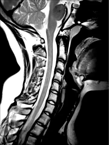
An MRI scan is often performed to diagnose disc damage and signs of compression of nerve roots. This diagnostic method has two major disadvantages. On the one hand, you only ever see one layer of the spine and never the entire construct in one picture; on the other hand, the patient is lying down. Sometimes the spine looks quite different when a patient is lying down versus standing up, which can be explained by differences in load distribution and corresponding biomechanics when the patient is standing up. Therefore, if you want to treat a patient using manual therapy techniques, a standing X-ray of the entire spine is far more helpful and gives a far more realistic picture than an MRI scan.
Source: Dr. Matthias Meier
In this case, a 17-year-old woman is complaining of chronic headaches. An MRI scan did not reveal any pathological changes other than a somewhat steep cervical spine and discrete disc bulges, thus no specific measures were recommended other than physiotherapy. Standing X-ray diagnostics, however, revealed a distinct kink between the fourth and fifth cervical vertebrae (arrow), indicating an injury to the so-called posterior longitudinal ligament and interrupting the harmonic curve of the posterior longitudinal edge. This disruption of the structure can cause the spinal cord, which is contained within the spinal column, to experience a change in tension. The patient’s reaction will always be to tense the neck muscles in an attempt to prevent this misalignment from becoming more severe to the extent possible. The kink is not visible via MRI, but is clearly visible on X-ray. The recommended treatment is clear based on the diagnosis that has now been established. The drawn curve illustrates the position the cervical spine should be in.
Source: Dr. Matthias Meier
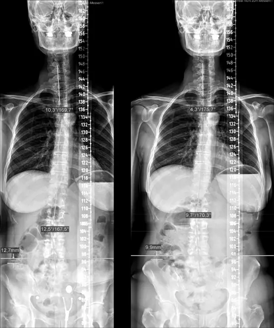
52-year-old lady with menstrual cramps, back pain, headaches, and cardiac arrhythmia. The left picture clearly shows that the head deviates to the left and a kink of 10.3° at the upper thoracic spine and a contrasting kink of 12.3° at the lumbar spine. A pelvic obliquity of 12.3 mm also interferes with the symmetry. After 29 treatments, the head was able to be centered and the respective values reduced to 4.3° at the upper thoracic spine and 9.7° at the lumbar spine. The pelvic obliquity was reduced to 9.9 mm. While this might not look too spectacular, it brings with it the disappearance of headaches, cardiac arrhythmia and menstrual cramps, as well as a significant reduction of the back pain.
Source: Dr. Matthias Meier
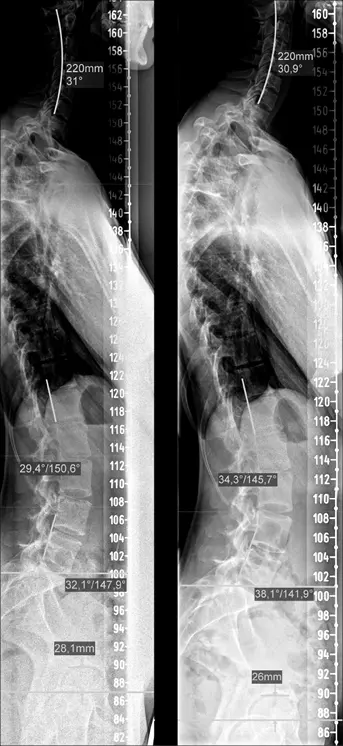
Young woman (17) suffering from headaches and scoliosis. In the left picture, you can see that the cervical spine no longer follows the curve at the upper segments, but falls forward (even if only slightly), the lumbar spine shows a curve of 29.4° (40° is considered physiological). After treatment, the cervical spine was harmonized (upper segments follow the curve) and the lumbar spine now shows 34.3°. The two images are separated by an interval of two weeks, in this time the young woman grew 1.5 cm.
Source: Dr. Matthias Meier
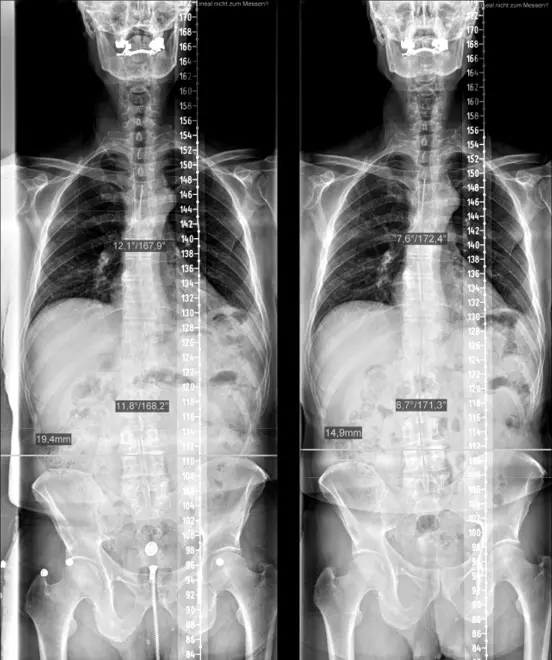
72-year-old man with prostate enlargement, elbow pain on both sides and foot pain on the right. After 29 treatments, the malpositions improved from 12.1° to 7.6° in the thoracic spine, from 11.8° to 8.7° in the lumbar spine, and the pelvic obliquity from 19.4 mm to 14.9 mm. The result is the difference between getting up six times each night to go to the bathroom with medication and getting up one to two times without medication and occasionally even being able to sleep through the night. Both his elbow and foot pain disappeared as well.
Source: Dr. Matthias Meier
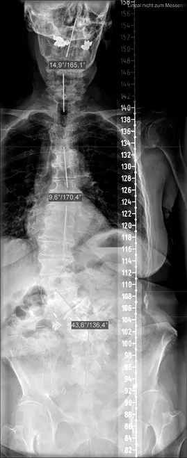
What about a spine like this? Do you think this elderly lady has normal blood pressure? Sleeps well? Has regular digestion? You guessed it—no, she doesn’t. The physical stress on the nervous system is clearly visible here. Organ dysfunctions and connective tissue instabilities of the knees with subsequent arthrosis on both sides have already occurred. Standing upright and walking becomes a torture and medication is taken to make the situation bearable. The cause can be clearly seen in the picture. However, the possibilities for correction of the biomechanics are significantly limited since the mobility of the individual vertebral segments has decreased significantly. Nonetheless, symptom relief by manipulation of the spinal components can still be achieved. Even if no structural change occurs, the patient can usually reduce the amount of medication needed.
It is clear, then, that the position of the spine determines pain, organ function as well as activity and dominance of the autonomic nervous system, and therefore may contribute to disease development. It is important to take this into account when treating patients with chronic diseases in order to be able to treat them causally.
There is another interesting aspect here. When the structure of the spine is reconstructed, old symptoms may sometimes temporarily reappear. An example of this would be a young woman who reported having cardiac arrhythmia indicated that disappeared after a few treatments, but after several weeks of therapy, she developed a feeling of instability in her right shoulder that she had not had for years. Five years ago, she had suffered a shoulder dislocation while serving in a tennis match, which she was able to “pop” back in spontaneously. Afterwards she had a feeling of instability for a while, which improved with physiotherapy and training. This feeling of instability came back after a certain number of therapies, but lasted only a few weeks. It is almost like turning back time with therapy and the patient is reliving their symptoms in reverse order. In other words, certain symptoms that occur as a result of a corresponding spinal misalignment may disappear as the misalignment continues, whereas others may be added that can be classified as more dangerous (cardiac arrhythmia vs. shoulder pain). The body tries to compensate as much as it can to maintain function, but gives clear signals when there is a serious problem.
Opioids
The opioid crisis in the U.S. dramatically illustrates how the central nervous system can be abused and what the consequences of doing so can be. CNN reports that in 2017, approximately 1.7 million Americans suffered from the effects of prescribed opioids and 652,000 people became addicted to heroin.2 Approximately 70,200 people died from substance overdoses, of which 47,600 were opioid overdoses. In 2016 and 2017, more than 130 people died each day from an opiate overdose (U.S. Department of Health & Human Services). In 2011, 240 billion milligrams of morphine were prescribed, then in 2017 it was “only” 171 billion milligrams.3 Globally, illicit drug sales, fueled in part by prescription drug abuse, are estimated at approximately $320 billion per year.
Читать дальшеИнтервал:
Закладка:
Похожие книги на «The Human Race – Too Smart to Survive»
Представляем Вашему вниманию похожие книги на «The Human Race – Too Smart to Survive» списком для выбора. Мы отобрали схожую по названию и смыслу литературу в надежде предоставить читателям больше вариантов отыскать новые, интересные, ещё непрочитанные произведения.


Обсуждение, отзывы о книге «The Human Race – Too Smart to Survive» и просто собственные мнения читателей. Оставьте ваши комментарии, напишите, что Вы думаете о произведении, его смысле или главных героях. Укажите что конкретно понравилось, а что нет, и почему Вы так считаете.