Ridley's The Vulva
Здесь есть возможность читать онлайн «Ridley's The Vulva» — ознакомительный отрывок электронной книги совершенно бесплатно, а после прочтения отрывка купить полную версию. В некоторых случаях можно слушать аудио, скачать через торрент в формате fb2 и присутствует краткое содержание. Жанр: unrecognised, на английском языке. Описание произведения, (предисловие) а так же отзывы посетителей доступны на портале библиотеки ЛибКат.
- Название:Ridley's The Vulva
- Автор:
- Жанр:
- Год:неизвестен
- ISBN:нет данных
- Рейтинг книги:3 / 5. Голосов: 1
-
Избранное:Добавить в избранное
- Отзывы:
-
Ваша оценка:
Ridley's The Vulva: краткое содержание, описание и аннотация
Предлагаем к чтению аннотацию, описание, краткое содержание или предисловие (зависит от того, что написал сам автор книги «Ridley's The Vulva»). Если вы не нашли необходимую информацию о книге — напишите в комментариях, мы постараемся отыскать её.
Ridley’s The Vulva
Ridley’s The Vulva
Ridley's The Vulva — читать онлайн ознакомительный отрывок
Ниже представлен текст книги, разбитый по страницам. Система сохранения места последней прочитанной страницы, позволяет с удобством читать онлайн бесплатно книгу «Ridley's The Vulva», без необходимости каждый раз заново искать на чём Вы остановились. Поставьте закладку, и сможете в любой момент перейти на страницу, на которой закончили чтение.
Интервал:
Закладка:
The primitive amniotic cavity develops at approximately 7–9 days post ovulation, and its floor forms the primary ectoderm ( Figure 1.2b). The primary endoderm is probably formed from cells originating from the ectoderm that migrate around the blastocoelic cavity and enclose the yolk sac. The ectoderm covering the floor of the amniotic cavity and the endoderm forming the roof of the yolk sac are in apposition, and therefore establish the bilaminar embryonic disc ( Figure 1.2c). A projection of the yolk sac endoderm into the extraembryonic mesoderm forms the allantoic diverticulum which identifies the caudal end of the bilaminar embryonic disc and the site of the body stalk ( Figure 1.2d).
The primitive streak ( Figure 1.3a) is formed and lies caudally in the midline of the embryonic disc. The primitive streak subsequently generates the intraembryonic mesoderm, which migrates through the bilaminar embryonic disc, in the plane between ectoderm and endoderm ( Figure 1.3b), converting it into a trilaminar disc. The disc remains bilaminar at the caudal and rostral ends. The caudal end forms the cloacal membrane.
Table 1.1 Carnegie stages in early embryogenesis.
| Carnegie stage | Days – post ovulation | Approximate size (mm) | Important events in genital & urological tract embryogenesis |
|---|---|---|---|
| 1 | 1 (week 1) | 0.1–0.15 | Point of fertilisation |
| 2 | 2–3 | 0.1–0.2 | |
| 3 | 4–5 | 0.1–0.2 | Blastocyst forms |
| 4 | 5–6 | 0.1–0.2 | Embeds in endometrium |
| 5 | 7–12 (week 2) | 0.1–0.2 | Allantoic diverticulum formed |
| 6 | 13–15 | 0.2 | Cloacal membrane formed |
| 7 | 15–17 (week 3) | 0.4 | |
| 8 | 17–19 | 1–1.5 | Primordial germ cells present |
| 9 | 19–21 | 1.5–2.5 | Hindgut formed and urogenital septum migrates caudally |
| 10 | 22–23 (week 4) | 2–3.5 | |
| 11 | 23–26 | 2.5–4.5 | Genital tubercle, cloacal folds, and genital swellings form |
| 12 | 26–30 | 3–5 | |
| 13 | 28–32 (week 5) | 4–6 | Urogenital septum divides hindgut into urogenital sinus and rectum |
| 14 | 31–35 | 5–7 | Labioscrotal folds fuse to form perineal body |
| 15 | 35–42 | 7–9 | Indifferent gonad |
| 16 | 37–42 (week 6) | 8–11 | Paramesonephric ducts appear. Trigone of bladder and posterior urethra formed |
| 17 | 42–44 | 11–14 | |
| 18 | 44–48 (week 7) | 13–17 | Embryonic testis develops in male |
| 19 | 48–51 | 16–18 | Bladder and urethra formed |
| 20 | 51–53 (week 8) | 18–22 | Ovary develops |
| 21 | 56–60 | 27–31 | |
| 22 | 54–56 | 23–28 | |
| 23 | 56–60 | 27–31 | External genital primordium developed, but still indeterminate sex |
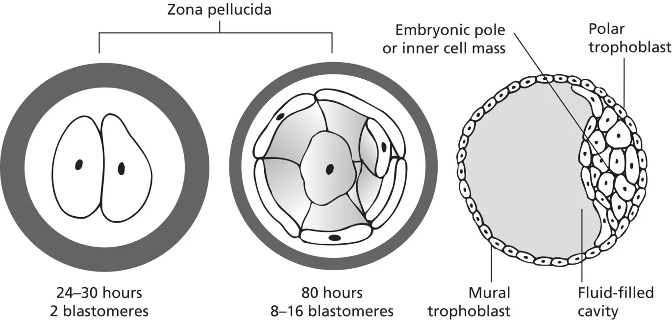
Figure 1.1 The conceptus is enclosed within an acellular envelope, the zona pellucida. After the formation of the blastocyst and dissolution of the zona pellucida, the characteristic fluid‐filled cavity, the embryonic pole or inner cell mass, and the mural trophoblast can be identified.
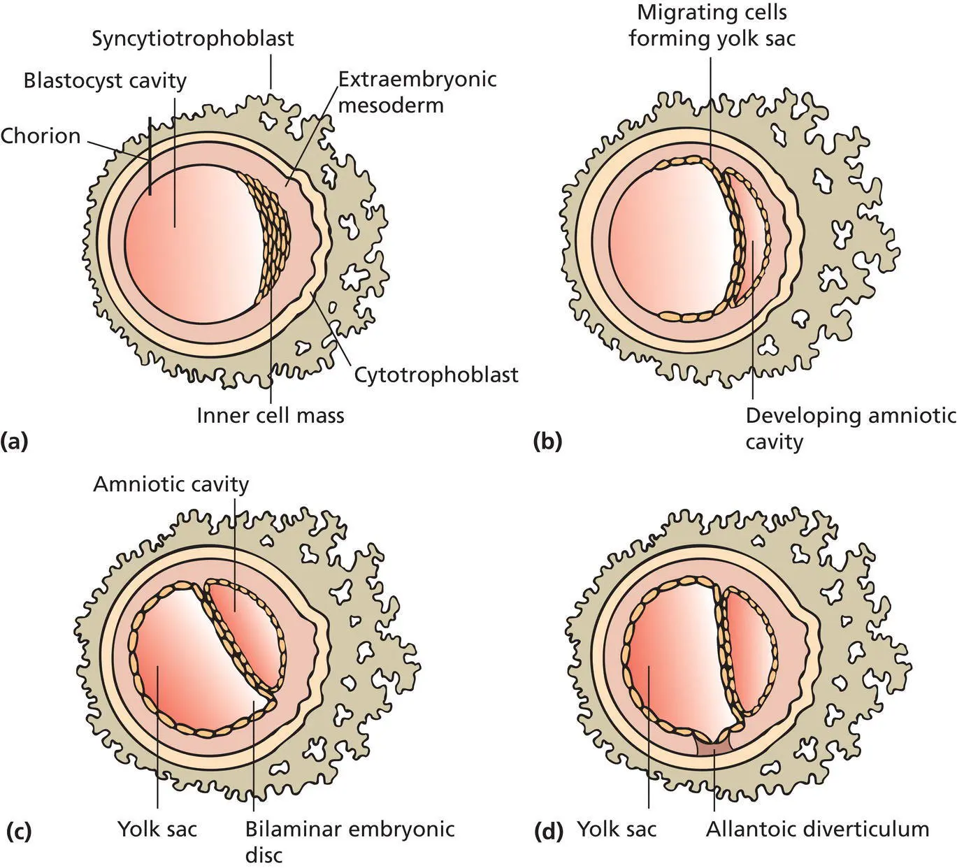
Figure 1.2 The conceptus continues to differentiate forming (a) the chorion, (b) the amniotic cavity, and (c) the yolk sac. The area of contact between the amniotic cavity and the yolk sac is the bilaminar embryonic disc. (d) Projection of the yolk sac endoderm into the mesoderm to form the allantoic diverticulum.
Carnegie stage 8
The primordial germ cells, which are the antecedents of the male and female gametes, are present in the endoderm around the allantoic diverticulum (now a ventral outpouching of the hind gut) and are usually seen in the 17–20 day embryo, although possible primordial germ cells have been identified at 13 days [8]. The primordial germ cells are ectodermal in origin, having migrated to the allantoic diverticulum from the epiblast, and they retain two functional X chromosomes in contrast to the somatic cells, which possess only one functional X chromosome [9]. From here, they migrate through the mesoderm surrounding the hindgut and into the dorsal mesentery. The final destination is the gonadal ridge, which they reach at about 35 days ( Figure 1.4).
Carnegie stage 9
The neural plate and the longitudinal neural ridges develop. The embryo flexes to accommodate the neural tube ( Figures 1.5a–c) and in so doing reorients the primitive embryonic tissues and their relationship to each other. The endoderm of the dorsal part of the yolk sac is drawn into the ventral concavity of the embryo and is subdivided into foregut, midgut, and hindgut ( Figure 1.6a,b). The hindgut appears about day 20 and is enclosed within the tail fold of the embryo. In this situation, the hindgut lies caudal to the rostral limit of the allantoic diverticulum and dorsal to the cloacal membrane. The mesoderm in the mid‐embryo region is divided into paraxial (surrounding the neural tube), lateral, and intermediate mesoderm. The intermediate mesoderm lies ventrally and lateral to the paraxial mesoderm, and differentiates medially into the gonadal ridge and laterally into the mesonephric region. The intermediate mesoderm at the rostral limit of the allantoic diverticulum extends dorsally and then caudally, in line with the curvature of the tail fold, dividing the hindgut into ventral and dorsal parts. As this division proceeds, the two parts of the hindgut remain in continuity with each other caudal to the advancing mesoderm of the urorectal septum. The caudal end of the hindgut is lined with endoderm and is known as the cloaca. On the ventral aspect of the cloaca there is a membrane which separates the endoderm from the surface ectoderm, the cloacal membrane . As development continues, a mesenchymal septum, the urogenital septum , migrates caudally ( Figures 1.7a–c).
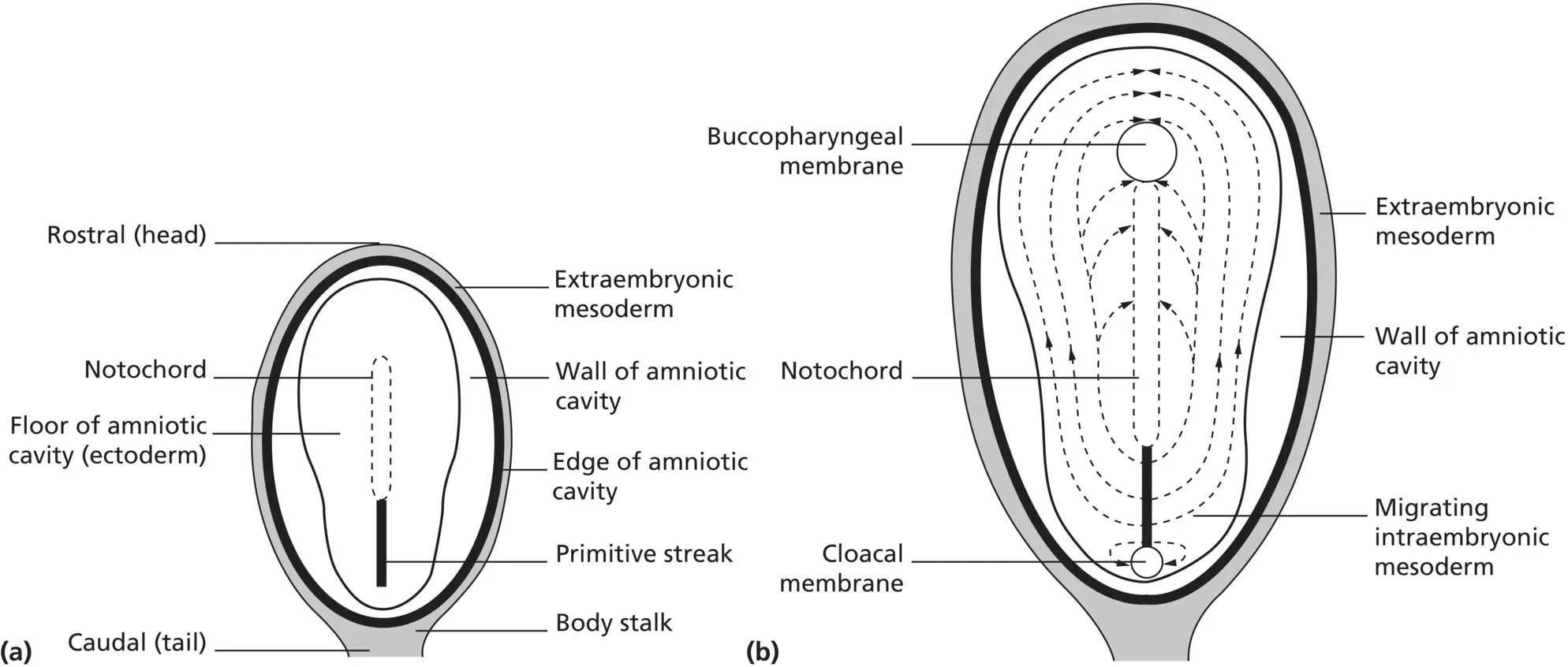
Figure 1.3 (a) The floor of the amniotic cavity, the dorsal surface of the bilaminar embryonic disc, revealing the primitive streak and notochord. (b) Intraembryonic mesoderm, generated by the primitive streak and interposed between the floor of the amniotic cavity and roof of the yolk sac, converts the bilaminar embryonic disc into a trilaminar disc. The buccopharyngeal and cloacal membranes remain bilaminar.
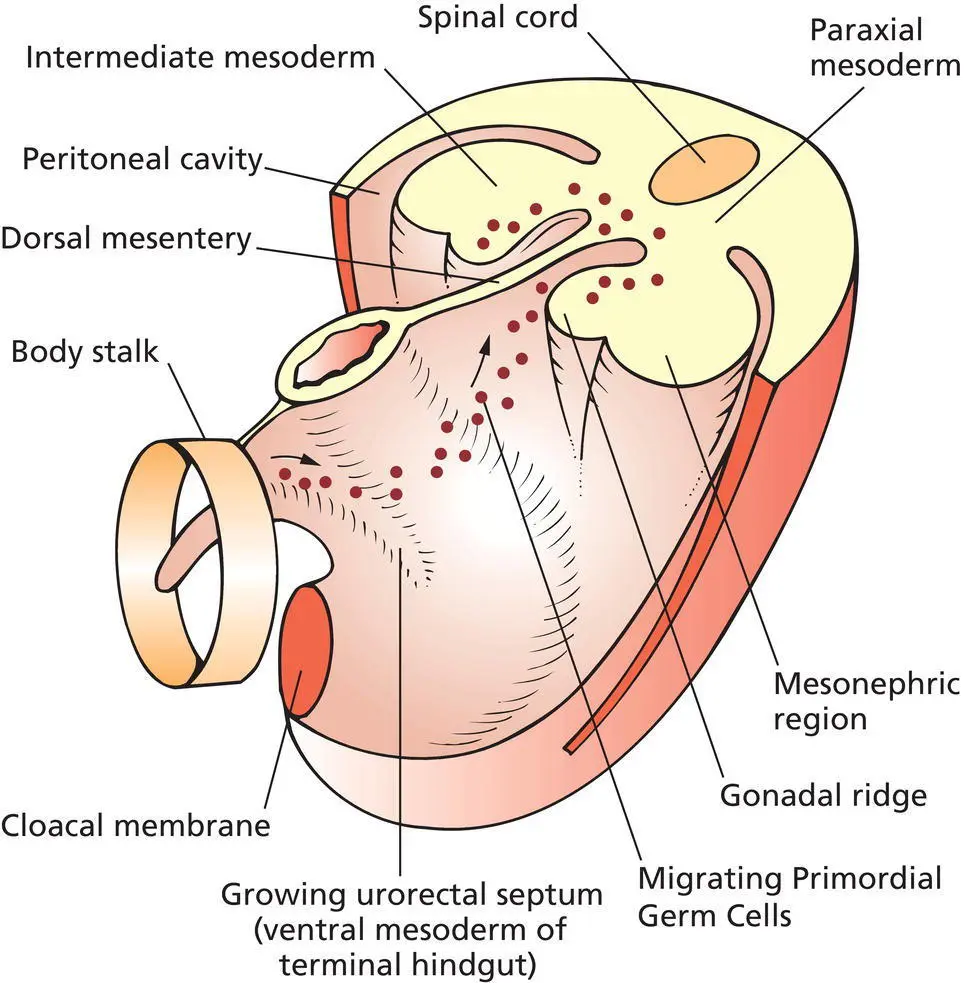
Figure 1.4 Migration of primordial germ cells into the genital ridge from the body stalk.
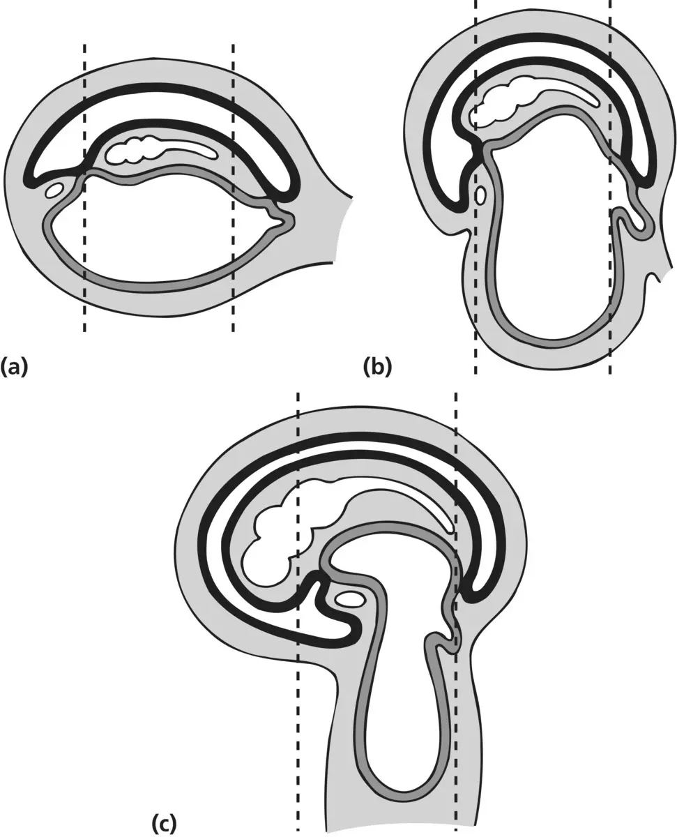
Figure 1.5 (a) As the neural tube is enclosed within the intraembryonic mesoderm, it lengthens and expands rostrally, causing a dorsal convexity and ventral concavity. (b) Further growth of the neural tube increases this curvature in the longitudinal plane (c) with the eventual formation of the head and tail folds.
Читать дальшеИнтервал:
Закладка:
Похожие книги на «Ridley's The Vulva»
Представляем Вашему вниманию похожие книги на «Ridley's The Vulva» списком для выбора. Мы отобрали схожую по названию и смыслу литературу в надежде предоставить читателям больше вариантов отыскать новые, интересные, ещё непрочитанные произведения.


Обсуждение, отзывы о книге «Ridley's The Vulva» и просто собственные мнения читателей. Оставьте ваши комментарии, напишите, что Вы думаете о произведении, его смысле или главных героях. Укажите что конкретно понравилось, а что нет, и почему Вы так считаете.