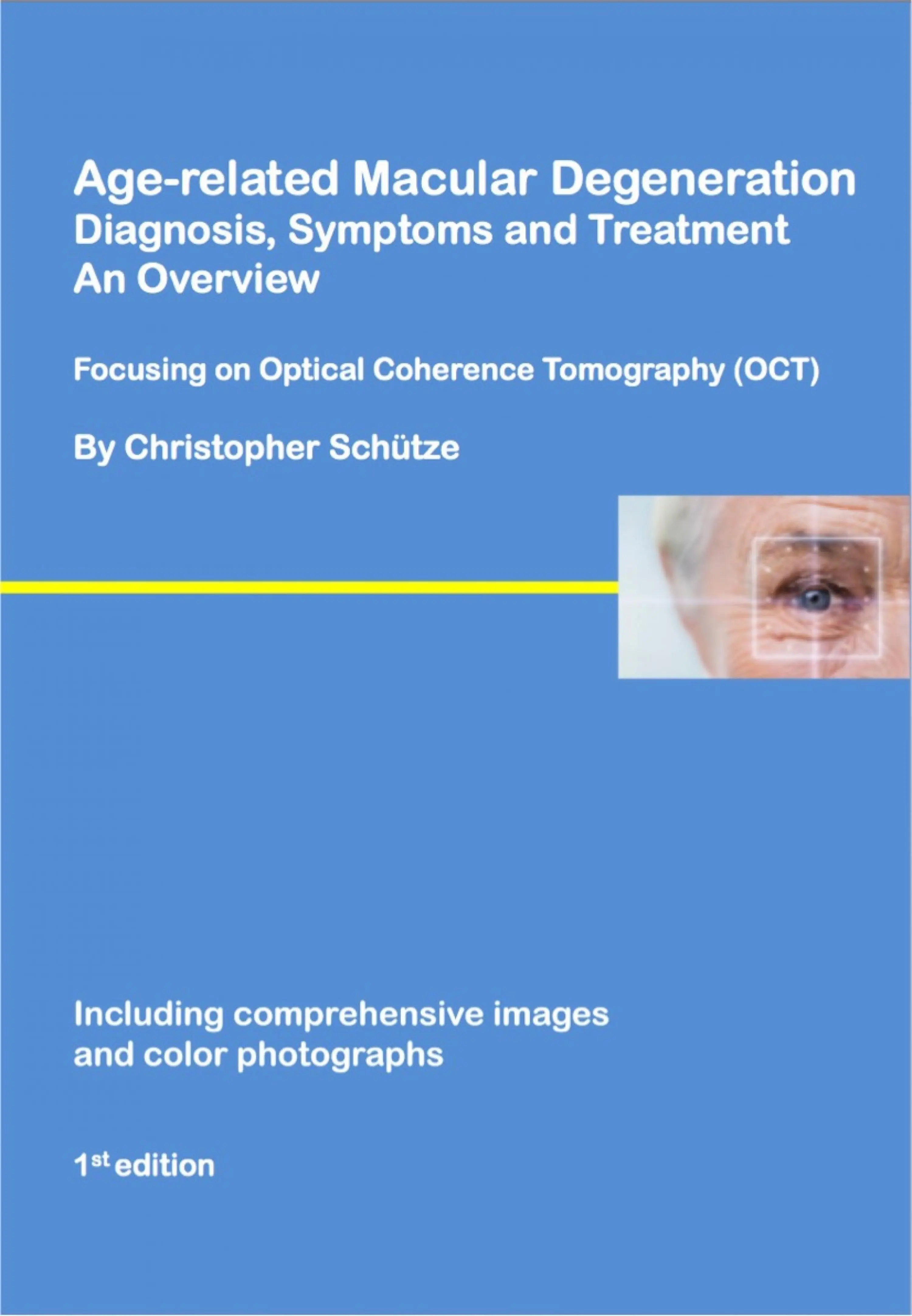
Christopher Schütze - Age-related macular degeneration
Здесь есть возможность читать онлайн «Christopher Schütze - Age-related macular degeneration» — ознакомительный отрывок электронной книги совершенно бесплатно, а после прочтения отрывка купить полную версию. В некоторых случаях можно слушать аудио, скачать через торрент в формате fb2 и присутствует краткое содержание. Жанр: unrecognised, на английском языке. Описание произведения, (предисловие) а так же отзывы посетителей доступны на портале библиотеки ЛибКат.
- Название:Age-related macular degeneration
- Автор:
- Жанр:
- Год:неизвестен
- ISBN:нет данных
- Рейтинг книги:3 / 5. Голосов: 1
-
Избранное:Добавить в избранное
- Отзывы:
-
Ваша оценка:
Age-related macular degeneration: краткое содержание, описание и аннотация
Предлагаем к чтению аннотацию, описание, краткое содержание или предисловие (зависит от того, что написал сам автор книги «Age-related macular degeneration»). Если вы не нашли необходимую информацию о книге — напишите в комментариях, мы постараемся отыскать её.
Wet AMD is marked by abnormal growth of blood vessels within the macular area (spot of sharpest sight) leading to fluid accumulation (edema) and retinal bleeding as well as scar formation. If the disease is not treated severe irreversible visual impairment will result. Treatment with new agents that are administered intravitreally and act against vascular endothelial growth factor (VEGF) are now available, impeding disease progression in many cases.
The present book gives a comprehensive overview about all aspects of AMD, useful for affected patients and interested readers and covers causes, epidemiological facts, diagnostics (emphasizing the topic optical coherence tomography-OCT) and treatment strategies of the disease, based on extensive literature research.
Age-related macular degeneration — читать онлайн ознакомительный отрывок
Ниже представлен текст книги, разбитый по страницам. Система сохранения места последней прочитанной страницы, позволяет с удобством читать онлайн бесплатно книгу «Age-related macular degeneration», без необходимости каждый раз заново искать на чём Вы остановились. Поставьте закладку, и сможете в любой момент перейти на страницу, на которой закончили чтение.
Интервал:
Закладка:
Chapter 2
Optical coherence tomography (OCT) 3:
2.1 Imaging the human retina by OCT in ophthalmology:
OCT is a non-invasive and non-contact imaging modality capable of generating cross-sectional images of the retina, the optic nerve, and the vitreous. Further, anterior segment OCT is capable of imaging anatomical structures of the anterior section of the eye (i.e. the cornea, the anterior chamber, or the lens). OCT is analogous to ultrasonography, however the technology uses light waves for imaging instead of sound waves. During an examination the eye is scanned transversely using a light beam. Cross-sectional intensity-based images can be generated and analyzed by means of a false color or gray scale image. Using this technology, the echo time delay and the amount of reflected or backscattered light is measured using low coherence interferometry (Kanski et al., 2008).
Indications for retinal OCT imaging:
- Diagnostics of pathological alterations in the macular area such as macular holes, central serous retinopathy, epiretinal membranes, macular edema, or vitreomacular traction (Hee et al., 1995b, Hee et al., 1995c, Hwang et al., 2012, Hee et al., 1995a, Tsunoda et al., 2012, Kanski et al., 2008).
- Documentation of disease progression and treatment response, i.e. retinal thickness measurements in neovascular AMD and its development during treatment follow-up (Sulzbacher et al., 2013, Golbaz et al., 2011, Kanski et al., 2008).
- Differentiation of retinal detachment of longer duration and retinoschisis (Kanski et al., 2008).
- Retinal nerve analysis and evaluation of retinal nerve fiber layer (RNFL) thickness, i.e. in patients with glaucoma (Chauhan et al., 2012, Kanski et al., 2008).
There are several color and gray scales used for representing intensity-based retinal images in OCT imaging. To mention only one example, images acquired by i.e. Cirrus SD-OCT (Cirrus HD-OCT; Carl. Zeiss Meditec, Dublin, California, USA) are displayed in the following way: light colors usually represent highly reflective layers (red and white), whereas retinal structures with low reflectivity are visualized in dark colors (blue and black). Structures with intermediate reflectivity are illustrated in green color. The plexiform and nerve fiber layer are visualized in red, yellow, or light green color.
Using Cirrus SD-OCT imaging as a representative example, the inner (IPL) and outer plexiform layers (OPL) are represented in light green color. The inner (INL) and outer nuclear layers (ONL) usually appear in blue or black color. The junction between the outer and inner segments of photoreceptors is displayed as a thin red structure in the outer retina. High-resolution OCT (i.e. Cirrus OCT) is further capable of differentiating retinal structures like the external limiting membrane (ELM) and the ganglion cell layer (GCL) (Kanski et al., 2008) (Fig.1).
Fig.1 shows an SD-OCT image of a healthy retina, displaying a cross-sectional image of retinal layers.
In essence, the bandwidth of the light source used in OCT imaging determines the axial resolution (Drexler, 2008). Enhanced axial OCT resolution can be achieved when using broad bandwidth light or light sources with a low coherence length (Drexler, 2008a). Image resolution of modern OCT machines used in clinical practice ranges between 5-8μm (Drexler, 2008a).
Конец ознакомительного фрагмента.
Текст предоставлен ООО «ЛитРес».
Прочитайте эту книгу целиком, купив полную легальную версию на ЛитРес.
Безопасно оплатить книгу можно банковской картой Visa, MasterCard, Maestro, со счета мобильного телефона, с платежного терминала, в салоне МТС или Связной, через PayPal, WebMoney, Яндекс.Деньги, QIWI Кошелек, бонусными картами или другим удобным Вам способом.
Интервал:
Закладка:
Похожие книги на «Age-related macular degeneration»
Представляем Вашему вниманию похожие книги на «Age-related macular degeneration» списком для выбора. Мы отобрали схожую по названию и смыслу литературу в надежде предоставить читателям больше вариантов отыскать новые, интересные, ещё непрочитанные произведения.


Обсуждение, отзывы о книге «Age-related macular degeneration» и просто собственные мнения читателей. Оставьте ваши комментарии, напишите, что Вы думаете о произведении, его смысле или главных героях. Укажите что конкретно понравилось, а что нет, и почему Вы так считаете.