Alister Hardy - The Open Sea - The World of Plankton
Здесь есть возможность читать онлайн «Alister Hardy - The Open Sea - The World of Plankton» — ознакомительный отрывок электронной книги совершенно бесплатно, а после прочтения отрывка купить полную версию. В некоторых случаях можно слушать аудио, скачать через торрент в формате fb2 и присутствует краткое содержание. Жанр: unrecognised, на английском языке. Описание произведения, (предисловие) а так же отзывы посетителей доступны на портале библиотеки ЛибКат.
- Название:The Open Sea: The World of Plankton
- Автор:
- Жанр:
- Год:неизвестен
- ISBN:нет данных
- Рейтинг книги:3 / 5. Голосов: 1
-
Избранное:Добавить в избранное
- Отзывы:
-
Ваша оценка:
The Open Sea: The World of Plankton: краткое содержание, описание и аннотация
Предлагаем к чтению аннотацию, описание, краткое содержание или предисловие (зависит от того, что написал сам автор книги «The Open Sea: The World of Plankton»). Если вы не нашли необходимую информацию о книге — напишите в комментариях, мы постараемся отыскать её.
The Open Sea: The World of Plankton — читать онлайн ознакомительный отрывок
Ниже представлен текст книги, разбитый по страницам. Система сохранения места последней прочитанной страницы, позволяет с удобством читать онлайн бесплатно книгу «The Open Sea: The World of Plankton», без необходимости каждый раз заново искать на чём Вы остановились. Поставьте закладку, и сможете в любой момент перейти на страницу, на которой закончили чтение.
Интервал:
Закладка:
FIG. 13
Diagrams showing the division of a simple pillbox-like type of diatom. a , a sketch of the cell before division has begun, b to d sections through the diatom after cell division to show stages in the formation of the new skeletal cell walls. Note that the upper cell in d is smaller than the original cell b.
Diatoms normally reproduce by simply dividing in two. The nucleus divides first and then the protoplasm becomes separated into two masses, each containing a nucleus, one at either end of the box; each mass of protoplasm now forms, between it and the other mass, a new valve as the halves of the pill-box are called. These new valves each fit closely their own part of the old box; we have in fact two pillboxes now instead of one, as is shown above in Fig. 13. They may separate entirely, or in some species they may remain attached to form long chains. It will be realised that in this process of repeated division by forming new half-boxes within the old, the average size of the diatoms so produced will tend to get smaller and smaller; at each division, as shown in the drawing, one of the new boxes will be the same size as the old one but the other must be smaller. Thus we find a considerable range in the size of diatoms of the same species, but there must be a limit to this reduction. After a certain number of such divisions there is formed what is called an auxospore, by which the original size is recovered; throwing off the old valves the cell becomes a bladder-like mass of protoplasm within which new valves are formed two or three times the size of the old discarded ones. Some diatoms seem to do this at definite seasons whereas others do so only at intervals of two or three years. By taking sample measurements of the diatoms forming some of the dense concentrations which are carried by currents about the North Sea, planktologists have been able to identify individual patches in their wanderings by the regular decrease in the size of their component cells (Wimpenny 1936; Lucas and Stubbings 1948). Some of these concentrations, as we shall see in Chapter 15have been thought to have a marked effect upon the herring fisheries; their wanderings may therefore be of economic interest.
In addition to forming auxospores, diatoms may produce what are called resting spores when conditions become adverse. The contents of the cell become concentrated in a central mass which forms a new thick wall of a different but characteristic shape and the old cell-wall is discarded. They now either sink into the deeper water layers or right to the bottom where they will remain till more suitable conditions return; thus some may pass the winter in a resting state and then come up to start active life again in the spring. Planktonic diatoms are not definitely known to have any sexual phase, but in some a number of smaller spores (microspores) have occasionally been observed to be formed within the cell-wall and it has been thought that these may be gametes (sex cells), but this is not yet established.
Not all diatoms are planktonic; in the shallow coastal regions there are numbers living on the bottom where sufficient light reaches it; these have much thicker shells than the more delicate floating forms and are more uniform in character. What makes the planktonic diatoms so interesting is the variety of devices that have been evolved to assist in their flotation. It is not the purpose of this book to attempt a systematic treatment of the groups of animals and plants. Here we shall just refer to some of the more important kinds of diatoms in relation to their mode of suspension. Dr. Marie Lebour’s excellent book (1930) on the planktonic forms should be studied for a full account.
Although so very small they have a considerable range in size; a few exceptionally large ones may be over one millimetre in diameter and the smallest may be but a few thousandths of this. Sketches of examples of the different genera to be mentioned are shown in Fig. 14and Plate 1, and some are also included in the photographs in Plate I. A number have the typical pill-box form, such as members of the genus Coscinodiscus and some of these are of comparatively large size; a few like C. concinnus may be just visible to the naked eye. These kinds have large vacuoles and would seem to be buoyed up by globules of oil. Members of this genus live singly, but others of allied genera may remain after division attached together to form long chains; the cells of Paralia and Guinardia are linked rigidly together by their valve surfaces, those of Thalassiosira form flexible chains as they are strung together by fine threads of protoplasm, and others like Lauderia are still more loosely held together by irregular strands of slime. The cells of Thalassiosira also produce such slime-strands but for a different purpose—around the margin of each valve (i.e. each ‘pill-box lid’) are a number of small hollow spines from which can be extruded long slender threads of slime so that they radiate on all sides like the strands of thistle-down and indeed act in the same way to assist in parachute-like support.
The remainder of the planktonic diatoms, while essentially built on this pillbox plan, have each valve or half-box modified into all sorts of shapes which increase their surface area in relation to their volume and so give greater frictional resistance to sinking. Some are flattened like thin sheets of paper and often twisted to some extent; these usually remain attached together to form long ribbons: such are Bellarochia, Eucampia and Streptotheca. Others are drawn out either into long thin hair-like forms such as Thalassiothrix longissima or into the more rigid pencil or needle-like members of the genus Rhizosolenia , pointed at each end; in the former the division plane between the two valves runs lengthwise along the thread whereas in the latter it occurs transversely to the long axis. Other forms again, such as Biddulphia and Corethron , increase their surface area by being provided with spines. This last method is developed to a remarkable extent by the many species of the genus Chaetoceros whose cells have four very long hair-like processes (two extending from each valve); long chains of these cells are formed and held together by their curving processes becoming interlocked with those of adjacent cells close against their point of origin.
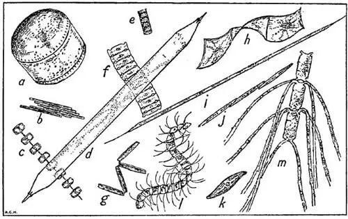
FIG. 14
Some characteristic plankton diatoms not shown in Plate I, all magnified ×90 diam. a , Coscinodiscus concinnus ; b , Bacillaria paradoxa ; c , Thalassiosira gravida ; d , Rhizosolenia styliformis ; e , Paralia sulcata ; f , Bellarochia maleus ; g , Thalassiothrix nitzschioides ; h , Streptotheca thamensis ; i , Rhizosolenia hebetata (form semispina ); j , Nitzschia seriata ; k , Gyrosigma sp. ; l , Chaetoceros curvisetus ; m , Ch. convolutus. The actual length represented by the longer side of this figure is 1/20th of an inch.
There are many species of Biddulphia in our waters, but one, B. sinensis ( Plate 1) is of special interest; it is now one of our commonest diatoms, often occurring in dense concentrations, yet it was unknown in European waters before 1903 when it was first recorded in the Heligoland Bight. It is a well known inhabitant of the coastal waters of the Indo-Pacific region, extending from the Red Sea to the coasts of China. It seems likely that it must have been brought, perhaps in ballast water, by some ship to the mouth of the Elbe; being tolerant of wide ranges of temperature and salinity, it found our waters congenial and spread rapidly. In the following years it was recorded further and further to the north until it reached a point a long way up the Norwegian coast where its further spread was probably checked by too cold water. More extraordinary, in view of the prevailing currents, was its spread down the Channel and into the Irish Sea; in the same year, 1909, it was reported for the first time both at Plymouth and off Port Erin in the Isle of Man. This would appear to provide evidence of an occasional reversal of the usual current flow up the Channel; indeed such a reversal has been suspected by some oceanographers on other grounds. Or was it transported to the western Channel and Irish Sea in the same way as it had apparently reached the Elbe? We may never know the answer to that. Some have maintained that Biddulphia sinensis must have been a native of our waters all the time and only just noticed at the beginning of the century; this, however, can hardly be so because of extensive collections that were made, particularly by the Kiel planktologists, throughout the eighteen-nineties.
Читать дальшеИнтервал:
Закладка:
Похожие книги на «The Open Sea: The World of Plankton»
Представляем Вашему вниманию похожие книги на «The Open Sea: The World of Plankton» списком для выбора. Мы отобрали схожую по названию и смыслу литературу в надежде предоставить читателям больше вариантов отыскать новые, интересные, ещё непрочитанные произведения.


Обсуждение, отзывы о книге «The Open Sea: The World of Plankton» и просто собственные мнения читателей. Оставьте ваши комментарии, напишите, что Вы думаете о произведении, его смысле или главных героях. Укажите что конкретно понравилось, а что нет, и почему Вы так считаете.