Jane Flint - Principles of Virology, Volume 1
Здесь есть возможность читать онлайн «Jane Flint - Principles of Virology, Volume 1» — ознакомительный отрывок электронной книги совершенно бесплатно, а после прочтения отрывка купить полную версию. В некоторых случаях можно слушать аудио, скачать через торрент в формате fb2 и присутствует краткое содержание. Жанр: unrecognised, на английском языке. Описание произведения, (предисловие) а так же отзывы посетителей доступны на портале библиотеки ЛибКат.

- Название:Principles of Virology, Volume 1
- Автор:
- Жанр:
- Год:неизвестен
- ISBN:нет данных
- Рейтинг книги:5 / 5. Голосов: 1
-
Избранное:Добавить в избранное
- Отзывы:
-
Ваша оценка:
Principles of Virology, Volume 1: краткое содержание, описание и аннотация
Предлагаем к чтению аннотацию, описание, краткое содержание или предисловие (зависит от того, что написал сам автор книги «Principles of Virology, Volume 1»). Если вы не нашли необходимую информацию о книге — напишите в комментариях, мы постараемся отыскать её.
Volume I: Molecular Biology
Volume II: Pathogenesis and Control
Principles of Virology, Fifth Edition
Principles of Virology, Volume 1 — читать онлайн ознакомительный отрывок
Ниже представлен текст книги, разбитый по страницам. Система сохранения места последней прочитанной страницы, позволяет с удобством читать онлайн бесплатно книгу «Principles of Virology, Volume 1», без необходимости каждый раз заново искать на чём Вы остановились. Поставьте закладку, и сможете в любой момент перейти на страницу, на которой закончили чтение.
Интервал:
Закладка:
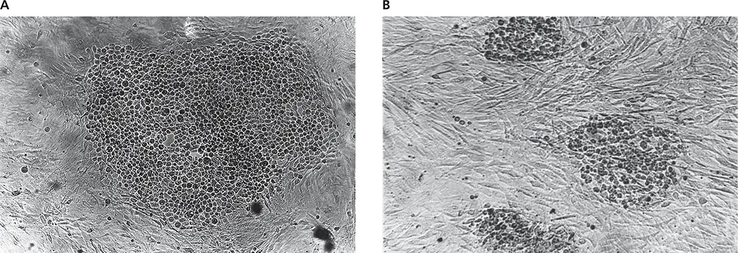
Figure 2.9 Transformation assay. Chicken cells transformed by two different strains of Rous sarcoma virus are shown. Loss of contact inhibition causes cells to pile up rather than grow as a monolayer. One focus is seen in panel Aand three foci are seen in panel Bat the same magnification. Courtesy of H. Hanafusa, Osaka Bioscience Institute.
BOX 2.6
METHODS
End-point dilution assays
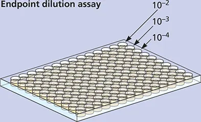
End-point dilution assays are usually carried out in multiwell plastic plates (see the figure above). In the example shown in the adjacent table above, 10 monolayer cell cultures were infected with each virus dilution. After the incubation period, plates that displayed cytopathic effect were scored +. At high dilutions, none of the cell cultures are infected because no infectious particles are delivered to the cells; at low dilutions, every culture is infected. The end pointis the dilution of virus that affects 50% of the test units. This number can be calculated from the data and expressed as 50% infectious dose (ID 50) per milliliter. Fifty percent of the cell cultures displayed cytopathic effect at the 10 –5dilution, and therefore, the virus stock contains 10 5TCID 50(tissue culture infectious dose) units.
| Virus dilution | Cytopathic effect | |||||||||
|---|---|---|---|---|---|---|---|---|---|---|
| 10 –2 | + | + | + | + | + | + | + | + | + | + |
| 10 –3 | + | + | + | + | + | + | + | + | + | + |
| 10 –4 | + | + | – | + | + | + | + | + | + | + |
| 10 –5 | – | + | + | – | + | – | – | + | – | + |
| 10 –6 | – | – | – | – | – | – | + | – | – | – |
| 10 –7 | – | – | – | – | – | – | – | – | – | – |
In most cases, the 50% end point does not fall on a dilution tested as shown in the example; for this reason, various statistical procedures have been developed to calculate the end point of the titration. In one popular method, the dilution containing the ID 50is identified by interpolation between the dilutions on either side of this value. The assumption is made that the location of the 50% end point varies linearly with the log of the dilution. Because the number of test units used at each dilution is usually small, the accuracy of this method is relatively low. For example, if six test units are used at each 10-fold dilution, differences in virus titer of only 50-fold or more can be detected reliably. The method is illustrated in the second example below, in which the lethality of poliovirus in mice is the end point. Eight mice were inoculated per dilution. In the method of Reed and Muench, the results are pooled, as shown in the table below, which equalizes chance variations (another way to achieve the same result would be to utilize greater numbers of animals at each dilution). The interpolated value of the 50% end point, which in this case falls between the 5th and 6th dilutions, is calculated to be 10 –6.5. The virus sample therefore contains 10 6.5LD 50(50% lethal dose). The LD 50may also be calculated as the concentration of the stock virus in PFU per milliliter (1 × 10 9) times the 50% end-point titer. In the example shown, the LD 50is 3 × 10 2PFU.
Reed LJ, Muench H. 1938. A simple method of estimating fifty percent endpoints. Am J Hyg 27:493–497.
| Dilution | Alive | Dead | Total alive | Total dead | Mortality ratio | Mortality (%) |
|---|---|---|---|---|---|---|
| 10 –2 | 0 | 8 | 0 | 40 | 0/40 | 100 |
| 10 –3 | 0 | 8 | 0 | 32 | 0/32 | 100 |
| 10 –4 | 1 | 7 | 1 | 24 | 1/25 | 96 |
| 10 –5 | 0 | 8 | 1 | 17 | 1/18 | 94 |
| 10 –6 | 2 | 6 | 3 | 9 | 3/12 | 75 |
| 10 –7 | 5 | 3 | 8 | 3 | 8/11 | 27 |
Although the linear nature of the dose-response curve indicates that a single particle is capable of initiating an infection (one-hit kinetics) ( Fig. 2.8), the high particle-to-PFU ratio of many viruses demonstrates that not all virus particles are successful. High values are sometimes caused by the presence of noninfectious particles with genomes that harbor lethal mutations or that have been damaged during growth or purification ( defective particles). An alternative explanation is that although all viruses in a preparation are in fact capable of initiating infection, not all of them succeed because of the complexity of the infectious cycle. Failure at any one step in the cycle prevents completion. In this case, a high particle-to-PFU ratio indicates not that most particles are defective but, rather, that they failed to complete the infection.
Table 2.1 Particle-to-PFU ratios of some animal viruses
| Virus | Particle/PFU ratio |
|---|---|
| Papillomaviridae | |
| Papillomavirus | 10,000 |
| Picornaviridae | |
| Poliovirus | 30–1,000 |
| Herpesviridae | |
| Herpes simplex virus | 50–200 |
| Polyomaviridae | |
| Polyomavirus | 38–50 |
| Simian virus 40 | 100–200 |
| Adenoviridae | 20–100 |
| Poxviridae | 1–100 |
| Orthomyxoviridae | |
| Influenza virus | 20–50 |
| Reoviridae | |
| Reovirus | 10 |
| Alphaviridae | |
| Semliki Forest virus | 1–2 |
Measurement of Virus Particles
Although the numbers of virus particles and infectious units are often not equal, assays for particle number are frequently used to approximate the number of infectious particles present in a sample. For example, assuming that the ratio of infectious units to physical particles is constant, the concentration of viral DNA or protein can be used to estimate the number of infectious particles. Biochemical or physical assays are usually more rapid and easier to carry out than those for infectivity, which may be slow, cumbersome, or impossible. Assays for subviral components also provide information on particle number if the amount of these components in each virus particle is known.
Electron Microscopy
With few exceptions, virus particles are too small to be observed directly by light microscopy. However, they can be seen readily in the electron microscope. If a sample contains only one type of virus, the particle count can be determined. A virus preparation is mixed with a known concentration of latex beads, and the numbers of virus particles and beads are then counted, allowing the concentration of the virus particles in the sample to be determined by comparison.
Читать дальшеИнтервал:
Закладка:
Похожие книги на «Principles of Virology, Volume 1»
Представляем Вашему вниманию похожие книги на «Principles of Virology, Volume 1» списком для выбора. Мы отобрали схожую по названию и смыслу литературу в надежде предоставить читателям больше вариантов отыскать новые, интересные, ещё непрочитанные произведения.


Обсуждение, отзывы о книге «Principles of Virology, Volume 1» и просто собственные мнения читателей. Оставьте ваши комментарии, напишите, что Вы думаете о произведении, его смысле или главных героях. Укажите что конкретно понравилось, а что нет, и почему Вы так считаете.