The Peripheral T-Cell Lymphomas
Здесь есть возможность читать онлайн «The Peripheral T-Cell Lymphomas» — ознакомительный отрывок электронной книги совершенно бесплатно, а после прочтения отрывка купить полную версию. В некоторых случаях можно слушать аудио, скачать через торрент в формате fb2 и присутствует краткое содержание. Жанр: unrecognised, на английском языке. Описание произведения, (предисловие) а так же отзывы посетителей доступны на портале библиотеки ЛибКат.
- Название:The Peripheral T-Cell Lymphomas
- Автор:
- Жанр:
- Год:неизвестен
- ISBN:нет данных
- Рейтинг книги:3 / 5. Голосов: 1
-
Избранное:Добавить в избранное
- Отзывы:
-
Ваша оценка:
The Peripheral T-Cell Lymphomas: краткое содержание, описание и аннотация
Предлагаем к чтению аннотацию, описание, краткое содержание или предисловие (зависит от того, что написал сам автор книги «The Peripheral T-Cell Lymphomas»). Если вы не нашли необходимую информацию о книге — напишите в комментариях, мы постараемся отыскать её.
The first book of its kind,
presents a far-reaching survey of this complex and rare group of blood cancers. Featuring contributions from thought-leaders concerned with all aspects of PTCL, this authoritative text covers biology, epidemiology, classification, approved and emerging drugs, molecular genetics, and more. Detailed clinical chapters address diagnosis, prognosis, and treatment of each of the major PTCL subtypes identified in the 2018 WHO Classification of Tumors of Hematopoietic and Lymphoid Tissues. This much-needed resource:
Covers the biological basis, epidemiology, classification, and treatment of PTCL Discusses the future of the field, including global collaboration efforts and novel approaches to PCTL Explores the role of biologics in PTCL and autologous and allogeneic stem-cell transplantation Offers new insights on molecular pathogenesis, innovative therapeutics, and novel drug combinations Features contributions from the Chairs The T-Cell Lymphoma Forum: the world’s largest meeting focused on PTCL Reflecting the unique epidemiology and genetic diversity of the PTCL,
is an indispensable source of data, insight, and references for the medical community, particularly those working in oncology and hematology.
The Peripheral T-Cell Lymphomas — читать онлайн ознакомительный отрывок
Ниже представлен текст книги, разбитый по страницам. Система сохранения места последней прочитанной страницы, позволяет с удобством читать онлайн бесплатно книгу «The Peripheral T-Cell Lymphomas», без необходимости каждый раз заново искать на чём Вы остановились. Поставьте закладку, и сможете в любой момент перейти на страницу, на которой закончили чтение.
Интервал:
Закладка:
Sc‐RNA‐seq has provided reference maps for circulating and tissue‐based T cells and their functional hierarchies [16]. Among relevant transcriptional differences between peripheral blood and tissue‐based T cells, cytoskeleton, cell–matrix interaction, and cell proliferation modules have emerged, underscoring the transcriptional imprint of tissue microenvironment. This information must be considered in the interpretation of gene expression profiling studies on PTCL, which have mostly relied on the differential comparison with normal T‐cell subsets from peripheral blood and/or cell lines.
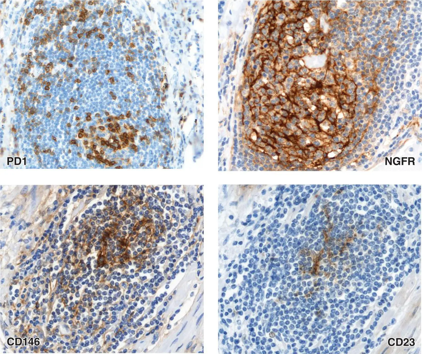
Figure 1.2 Representative immunohistochemical staining for PD1 (upper left) and NGFR/CD271 (upper right) highlighting the presence of PD1+ T follicular helper cells and of a formed meshwork of follicular dendritic cells within a tertiary lymphoid follicle in a case of non‐small‐cell lung carcinoma. Immunohistochemical staining for CD146 (lower left) and CD23 (lower right) highlighting the early branching of perivascular CD146+ mesenchymal elements that partially display CD23 expression as a marker of follicular dendritic cell differentiation in the same non‐small‐cell lung carcinoma.
Source: Claudio Tripodo, Stefano Pilleri.
Moreover, at the Sc‐RNA‐seq level, a clear dichotomy in the CD8+ effector T‐cell cluster emerges as conserved across tissue sites, based on the predominance of cytotoxicity‐related genes or cytokine and chemokine genes [16].
Tissue‐conserved Sc‐RNA‐seq transcriptional clustering also identifies distinct Treg, CD4+ naïve/central memory resting and CD4+/CD8+ resting clusters, IFN response, and proliferation gene clusters characterizing different CD4+ T‐cell activation states [16]. Interestingly, hallmarks of such transcriptional modules feature the concomitant expression of genes coding for transcription factors involved in divergent‐fate specifications (e.g. FOXP3, IRF4, and TOX2), highlighting the dynamical regulation of promiscuous transcription factors [17, 18]. Again, this concept may represent a relevant note of caution in the interpretation of transcription factor expression data in PTCL clonal populations as hallmarks of stable clone differentiative/phenotypic states.
The expression and activity of specific transcription factors involved in fate specification, such as T‐bet and IRF4, is bound to that of the pleiotropic transcription factor MYC in enabling TCR‐driven immune function through the regulation of T‐cell proteomic and metabolic landscape [19]. MYC proficiency has been demonstrated to be required for adapting protein expression to TCR engagement in CD4+ and CD8+ cells. Through the regulation of amino acid transporter expression, MYC impacts on translational activity and on the associated increase of effector T‐cell biomass ( Figure 1.3) [19], which relates with the higher Myc expression levels in CD8+ T cells.
Moreover, upon T‐cell activation, MYC controls lactate transporter expression, regulating the feedback on glycolytic flux fueling T‐cell synthetic programs and proliferative attitude [19]. In PTCL‐NOS not otherwise specified, overexpression of MYC and its transcriptional targets involved in cell proliferation has been implicated in the worse prognosis of GATA3‐positive cases [20]. Nevertheless, in the light of the profound effects on the metabolic adaptation to TCR sensing, MYC may represent an underrated determinant of inter/intraclonal competition [21] underlying PTCL progression.
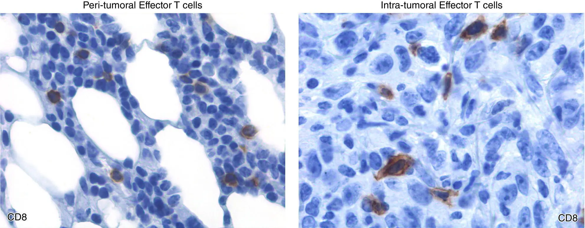
Figure 1.3 The biomass of CD8+ effector T cells is dramatically different between peritumoral (left panel) and intratumoral (right panel) compartments of a high‐grade carcinoma. Intra‐tumoral CD8+ effectors corresponding to an activated state showing a larger size.
Source: Claudio Tripodo, Stefano Pilleri.
This introductory chapter provides an updated view of the mature T‐cell constellation, in a less static view, as compared with canonical ontogeny‐focused outlines, providing information on the biological foundations of functional plasticity, phenotypical heterogeneity, and transcriptional dynamics that are recapitulated and magnified in peripheral T‐cell malignancies. It is imperative to recognize that the diversity, and complex function of normal T cells, is central to appreciating the many histologic and genetic subtypes of PTCL, and may aid in explaining the diverse behavior and presentation of these challenging and heterogeneous diseases.
Must Reads
Litman, G.W., Cannon, J.P., and Dinshaw, L.J.. (2005). Reconstructing immune phylogeny: new perspectives. Nat Rev Immunol 5(11): 866–879.
Restifo, N.P. and Gattinoni, L. (2013). Lineage relationship of effector and memory T‐cell. Curr Opin Immunol 25(5): 556–563.
Withers, D.R. (2016). Innate lymphoid cells regulation of adaptive immunity. Immunology 149(2): 123–130.
References
1 1 Swerdlow, S.H., Campo, E., Harris, N.L. et al. (2017). WHO Classification of Tumours of Haematopoietic and Lymphoid Tissues. Revised 4e. Lyon: IARC.
2 2 Kanhere, A., Hertweck, A., Bhatia, U. et al. (2012). T‐bet and GATA3 orchestrate Th1 and Th2 differentiation through lineage‐specific targeting of distal regulatory elements. Nat Commun 3: 1268.
3 3 Yang, X.O., Pappu, B.P., Nurieva, R. et al. (2008). T helper 17 lineage differentiation is programmed by orphan nuclear receptors RORα and RORγ. Immunity 28 (1): 29–39.
4 4 Plank, M.W., Kaiko, G.E., Maltby, S. et al. (2017). Th22 cells form a distinct Th lineage from Th17 cells in vitro with unique transcriptional properties and Tbet‐dependent Th1 plasticity. J Immunol 198 (5): 2182–2190.
5 5 Malik, S., Sadhu, S., Elesa, S. et al. (2017). Transcription factor Foxo1 is essential for IL‐9 induction in T helper cells. Nat Commun 8 (1): 815.
6 6 Xu, W., Zhao, X., Wang, X. et al. (2019). The transcription factor Tox2 drives T follicular helper cell development via regulating chromatin accessibility. Immunity 51 (5): 826–839.
7 7 Chen, W.J., Jin, W., Hardegen, N. et al. (2003). Conversion of peripheral CD4+CD25− naive T cells to CD4+CD25+ regulatory T cells by TGF‐β induction of transcription factor Foxp3. J Exp Med 198 (12): 1875–1886.
8 8 Tripathi, S.K. and Lahesmaa, R. (2014). Transcriptional and epigenetic regulation of T‐helper lineage specification. Immunol Rev 261 (1): 62–83.
9 9 Vahedi, G., Takahashi, H., Nakayamada, S. et al. (2012). STATs shape the active enhancer landscape of T cell populations. Cell 151 (5): 981–993.
10 10 Klose, C.S.N. and Artis, D. (2016). Innate lymphoid cells as regulators of immunity, inflammation and tissue homeostasis. Nat Immunol 17: 765–774.
11 11 Fousteri, G. and Zhu, J. (2018). T follicular helper‐like cells in inflamed non‐lymphoid tissues. Front Immunol 9: 1707.
12 12 Tripodo, C., Gri, G., and Piccaluga, P.P. (2010). Mast cells and Th17 cells contribute to the lymphoma‐associated pro‐inflammatory microenvironment of angioimmunoblastic T‐cell lymphoma. Am J Pathol 117 (2): 792–802.
13 13 Pepe, G., Di Napoli, A., Cippitelli, C. et al. (2018). Reduced lymphotoxin‐beta production by tumour cells is associated with loss of follicular dendritic cell phenotype and diffuse growth in follicular lymphoma. J Pathol 4 (2): 124–134.
14 14 Bilate, A.M., Bousbaine, D., Mesin, L. et al. (2016). Tissue‐specific emergence of regulatory and intraepithelial T cells from a clonal T cell precursor. Sci Immunol 1 (2): eaaf7471.
Читать дальшеИнтервал:
Закладка:
Похожие книги на «The Peripheral T-Cell Lymphomas»
Представляем Вашему вниманию похожие книги на «The Peripheral T-Cell Lymphomas» списком для выбора. Мы отобрали схожую по названию и смыслу литературу в надежде предоставить читателям больше вариантов отыскать новые, интересные, ещё непрочитанные произведения.


Обсуждение, отзывы о книге «The Peripheral T-Cell Lymphomas» и просто собственные мнения читателей. Оставьте ваши комментарии, напишите, что Вы думаете о произведении, его смысле или главных героях. Укажите что конкретно понравилось, а что нет, и почему Вы так считаете.