Mark W. Leitman - Manual for Eye Examination and Diagnosis
Здесь есть возможность читать онлайн «Mark W. Leitman - Manual for Eye Examination and Diagnosis» — ознакомительный отрывок электронной книги совершенно бесплатно, а после прочтения отрывка купить полную версию. В некоторых случаях можно слушать аудио, скачать через торрент в формате fb2 и присутствует краткое содержание. Жанр: unrecognised, на английском языке. Описание произведения, (предисловие) а так же отзывы посетителей доступны на портале библиотеки ЛибКат.
- Название:Manual for Eye Examination and Diagnosis
- Автор:
- Жанр:
- Год:неизвестен
- ISBN:нет данных
- Рейтинг книги:3 / 5. Голосов: 1
-
Избранное:Добавить в избранное
- Отзывы:
-
Ваша оценка:
Manual for Eye Examination and Diagnosis: краткое содержание, описание и аннотация
Предлагаем к чтению аннотацию, описание, краткое содержание или предисловие (зависит от того, что написал сам автор книги «Manual for Eye Examination and Diagnosis»). Если вы не нашли необходимую информацию о книге — напишите в комментариях, мы постараемся отыскать её.
is the leading introductory clinical textbook in the field, providing concise and practical coverage of anatomy, instrumentation, differential diagnosis and treatment of ophthalmic diseases and disorders. This accessible resource offers clear explanations of current diagnostic techniques, equipment and best practice; hundreds of full-color clinical photographs and diagrams; and step-by-step guidance on a range of key procedures. Author Mark Leitman, a practicing Ophthalmology specialist with nearly five decades’ experience, provides the foundational knowledge required to excel on an ophthalmology rotation. This is a must-have companion for medical students and junior doctors, trainee ophthalmologists and optometrists, optometry nurses, and ophthalmic technicians.
Manual for Eye Examination and Diagnosis — читать онлайн ознакомительный отрывок
Ниже представлен текст книги, разбитый по страницам. Система сохранения места последней прочитанной страницы, позволяет с удобством читать онлайн бесплатно книгу «Manual for Eye Examination and Diagnosis», без необходимости каждый раз заново искать на чём Вы остановились. Поставьте закладку, и сможете в любой момент перейти на страницу, на которой закончили чтение.
Интервал:
Закладка:
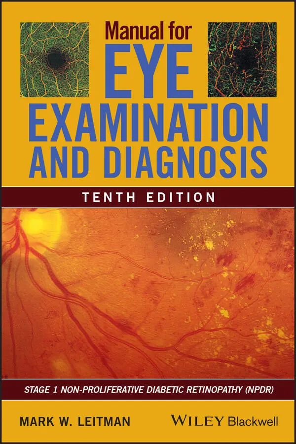
3 Retinopathy due to microvascular disease (see front and back cover) may result in macular edema. It is the primary reason for blindness before age 65. Patients with diabetes should have annual eye exams, because early treatment is critical. Retinopathy is rare in children before age 15.
Autoimmune (Graves’) thyroid disease
This is a condition in which an orbitopathy may be present with hyper‐ but also hypo‐ or euthyroid disease.
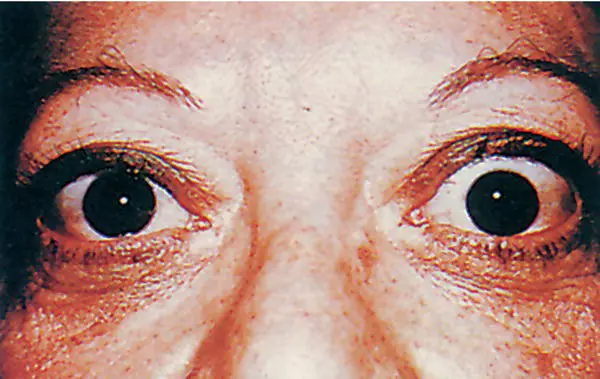
Fig 1 Thyroid exophthalmos with exposed sclera at superior limbus, due to bulging eye.
1 It is the most common cause of bulging eyes, referred to as exophthalmos (proptosis). This is due to fibroblast proliferation and mucopolysaccharide infiltration of the orbit. A small white area of sclera appearing between the lid and upper cornea is diagnostic of thyroid disease 90% of the time ( Fig. 1). This exposed sclera may be a result of exophthalmos or thyroid lid retraction due to the stimulation of Müller’s muscle that elevates the lid. Severe orbitopathy may be treated with radiation, surgical decompression of the orbit ( Fig. 3), or by administering steroids orally, intravenously, or by injection into the orbit. In January 2020, the FDA approved the human monoclonal antibody, teprotumumab, to treat proptosis, strabismus, and compressive optic neuropathy from thyroid eye disease. It reduced proptosis by more than 2 mm in 71–83% of patients.
1 Infiltration of eye muscles may cause diplopia, which is confirmed by a computed tomography (CT) scan ( Figs 2and 3).
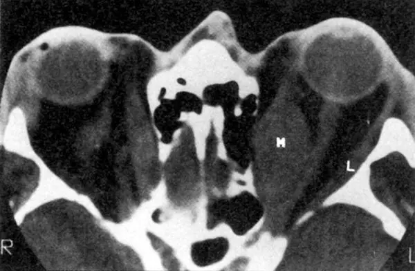
Fig 2 CT scan of thyroid orbitopathy showing fluid infiltration of the medial rectus muscle (M) and normal lateral rectus muscle (L) and proptosis. Compression of left optic nerve could cause optic neuropathy. This is called crowded apex syndrome .
Source : Courtesy of Jack Rootman.
1 Exophthalmos may cause excessive exposure of the eye in the day and an inability to close the lids at night (lagophthalmos), resulting in corneal dessication. Fig 3 Orbital CT scan of Graves’ orbitopathy before surgical decompression (above) and right orbital floor osteotomy (below). Often three, but rarely all four, bony walls may be opened. Note thickened extra ocular muscles.Source: Courtesy of Lelio Baldeschi, MD, and Ophthalmology, July 2007, Vol. 114, pp. 1395–1402.
2 Optic nerve compression is the worst complication and occurs in 4% of patients with thyroid disease. It could cause permanent loss of vision ( Fig. 2) and immediate intravenous steroids should be considered when vision is threatened.
Medications (ocular side effects)
Record patient medications. Those taking the following commonly prescribed drugs are often referred to an eye doctor to monitor ocular side effects.
Hydroxychloroquine (Plaquenil), initially used to treat malaria, is now a cornerstone medication used to treat autoimmune diseases, such as rheumatoid arthritis, lupus erythematosus, and Sjögren’s syndrome. It may cause retinal maculopathy with pigment changes resembling a “bull’s eye” ( Fig. 4). Patients should get a baseline eye exam before starting medication. It includes visual acuity, Amsler grid, color vision, and examination of the retina to rule out preexisiting maculopathy. The patient should follow up every 6 months. Depending on the dosage and the chronicity of use, the eye doctor will determine if additional tests are necessary. Risk increases if dosage exceeds 5 mg/kg. Toxicity is also related to the cumulative amount of the drug with 1% occurrence in the first 5 years and 2% after 10 years and if there is coexisting macular degeneration. These high‐dose chronically treated patients may also have routine monitoring of their visual fields and optical coherence tomography (OCT) testing (Figs 463 and 464).
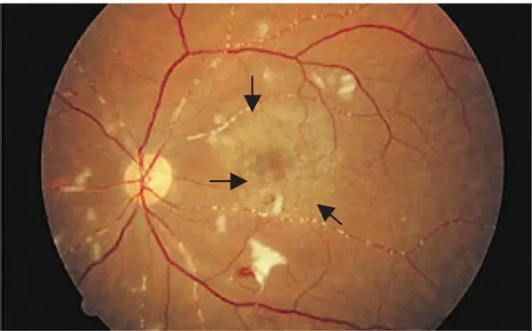
Fig 4 Bull’s eye maculopathy due to hydroxychloroquine in a patient with systemic lupus. The vasculitis and white cotton‐wool spots are due to lupus.
Source : Courtesy of Russel Rand, MD, and Arch. Ophthalmol ., Apr. 2000, Vol. 118, pp. 588–589. Copyright 2000, American Medical Association. All rights reserved.
The retina is also adversely affected by phenothiazine tranquilizers ( Fig. 5); niacin, a lipid‐lowering agent and interferon, used to treat multiple sclerosis and hepatitis C ( Figs 6– 8).
Ethambutol, rifampin, isoniazid, and streptomycin—taken mainly for tuberculosis—may all cause optic neuropathy. The antidepressants Paxil, Prozac, and Zoloft may also cause optic neuropathy. Corticosteroids may cause posterior subcapsular cataracts (Fig. 429), glaucoma, and a reduction in immunity that may increase the incidence of herpes virus and other infections.
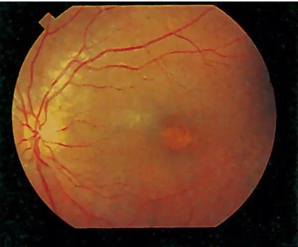
Fig 5 Phenothiazine maculopathy with pigment mottling of the macula.
Flomax (tamsulosin), the most common treatment for an enlarged prostate gland, increases the complications in cataract surgery by decreasing the ability to dilate the pupil, a condition referred to as intraoperative floppy iris syndrome (IFIS). Pupillary expansion devices ( Fig. 9) and additional pupillary dilating medications usually prevent complications.
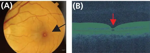
Fig 6 Tamoxifen maculopathy with crystalline deposits (A); and (B) OCT showing crystals in the fovea.
Source : Courtesy of Joao Liporaci, MD.
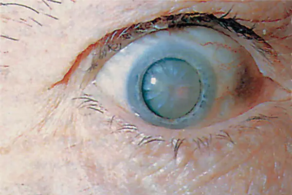
Fig 7 Tamoxifen causes cataracts.
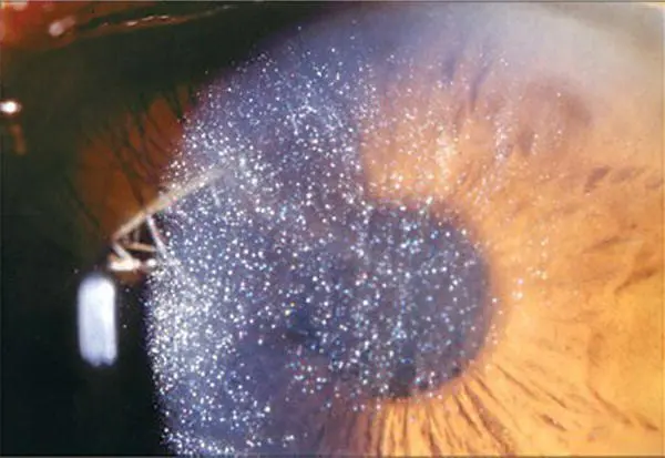
Fig 8 Besides causing maculopathy and cataracts, tamoxifen also causes crystal deposition in the cornea (keratopathy).
Source : Courtesy of Olga Zinchuk, MD, and Arch. Ophthalmol ., July 2006, Vol. 124, p. 1046. Copyright 2006, American Medical Association. All rights reserved.
Stevens–Johnson syndrome ( Fig. 10) is an immunologic reaction to a foreign substance, usually drugs, and most commonly sulfonamides, barbiturates, and penicillin. Some 100 other medications have also been implicated. It often affects the skin and mucous membranes. It could be fatal in 35% of cases.
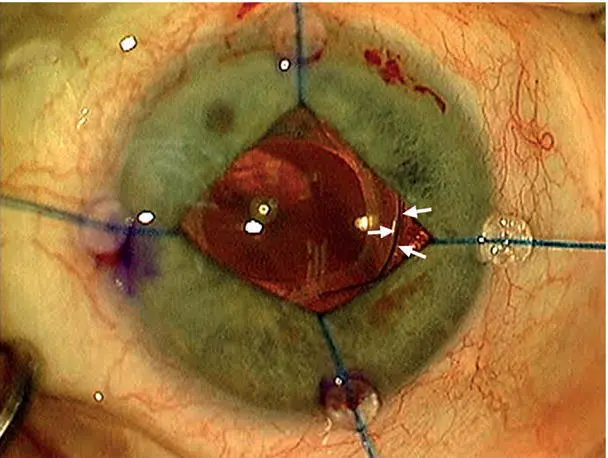
Fig 9 Iris retractors are one method used to open poorly dilated pupils during cataract surgery. Note edge of lens implant (↑) behind iris.
Читать дальшеИнтервал:
Закладка:
Похожие книги на «Manual for Eye Examination and Diagnosis»
Представляем Вашему вниманию похожие книги на «Manual for Eye Examination and Diagnosis» списком для выбора. Мы отобрали схожую по названию и смыслу литературу в надежде предоставить читателям больше вариантов отыскать новые, интересные, ещё непрочитанные произведения.


Обсуждение, отзывы о книге «Manual for Eye Examination and Diagnosis» и просто собственные мнения читателей. Оставьте ваши комментарии, напишите, что Вы думаете о произведении, его смысле или главных героях. Укажите что конкретно понравилось, а что нет, и почему Вы так считаете.