Louis Hardan - Protocols for Mobile Dental Photography with Auxiliary Lighting
Здесь есть возможность читать онлайн «Louis Hardan - Protocols for Mobile Dental Photography with Auxiliary Lighting» — ознакомительный отрывок электронной книги совершенно бесплатно, а после прочтения отрывка купить полную версию. В некоторых случаях можно слушать аудио, скачать через торрент в формате fb2 и присутствует краткое содержание. Жанр: unrecognised, на английском языке. Описание произведения, (предисловие) а так же отзывы посетителей доступны на портале библиотеки ЛибКат.
- Название:Protocols for Mobile Dental Photography with Auxiliary Lighting
- Автор:
- Жанр:
- Год:неизвестен
- ISBN:нет данных
- Рейтинг книги:4 / 5. Голосов: 1
-
Избранное:Добавить в избранное
- Отзывы:
-
Ваша оценка:
Protocols for Mobile Dental Photography with Auxiliary Lighting: краткое содержание, описание и аннотация
Предлагаем к чтению аннотацию, описание, краткое содержание или предисловие (зависит от того, что написал сам автор книги «Protocols for Mobile Dental Photography with Auxiliary Lighting»). Если вы не нашли необходимую информацию о книге — напишите в комментариях, мы постараемся отыскать её.
Protocols for Mobile Dental Photography with Auxiliary Lighting — читать онлайн ознакомительный отрывок
Ниже представлен текст книги, разбитый по страницам. Система сохранения места последней прочитанной страницы, позволяет с удобством читать онлайн бесплатно книгу «Protocols for Mobile Dental Photography with Auxiliary Lighting», без необходимости каждый раз заново искать на чём Вы остановились. Поставьте закладку, и сможете в любой момент перейти на страницу, на которой закончили чтение.
Интервал:
Закладка:
Treatment Planning
From the patient’s first visit to the dental clinic, dental photography can prove its worth as a diagnostic and evaluative tool. A complete set of oral photographs is essential to create a thorough treatment plan. It can be a very useful implement for the analysis of facial profiles, for the evaluation of prosthetic rehabilitation, for the detection of caries or enamel defects, and for the assessment of gingival health and periodontal pocket or ridge morphology prior to implant placement. Moreover, photographs can be sent to external specialists for a second opinion without need for patient consultation, facilitating better diagnosis and optimized treatment planning. For esthetic cases, photographs can be used to build a smile design so the patient can see the final outcome via a virtual mockup on screen that can be printed and transferred to the mouth with bis-acryl material (Figs 1 and 2). Many smile design softwares are evolving to be used on smartphones as an app or on a website where you can upload photographs directly from the image gallery.


FIGS 1 & 2The patient can see his final smile design before beginning treatment. The photographs serve to build this smile, and if the patient approves the design, the dentist can begin the treatment in a guided way.
Documentation and Self-Evaluation
For many years documentation in dental records relied on handwritten notes, radiographs, study models, and clinical photographs. Today most of this documentation is digitized for accessibility purposes. Photographs constitute a very effective archiving tool, providing improved documentation of clinical conditions over time and facilitating observation and monitoring.
Photographs also allow a more precise assessment of the completed work and the procedures followed, enabling effective self-evaluation. Details imperceptible to the human eye without magnification all of a sudden become clear. Moreover, the practitioner can easily examine and compare different procedures for different patients to determine which led to a better outcome, potentially improving future treatment planning.
With the evolution of smartphone cameras, accessibility is further enhanced because every dentist has a phone in his or her pocket that is designed to be user friendly. Photographs taken on a smartphone can be organized into folders by case and later evaluated on a larger screen to evaluate the quality of the work in hopes of improving it in future cases (Figs 3 and 4).
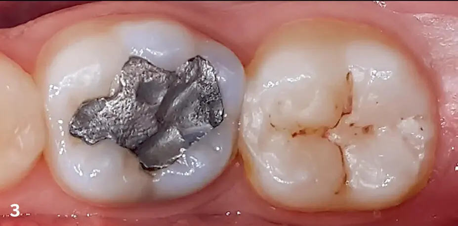
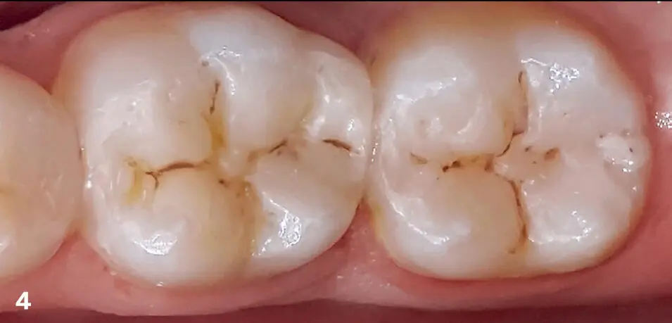
FIGS 3 & 4Before and 4 weeks after restoration of the first molar (and 1 year after restoration of the second molar). Examination of these photographs allows better assessment of the work performed than direct examination in the mouth and exposes any problems that can be corrected in the future.
Communication with the Patient and the Dental Technician
Effective communication between the practitioner and the patient is crucial for the success of any dental treatment, and a photograph is the simplest way to communicate dental information to patients. Images leave an impression on the patient and give him or her the sufficient confidence to move forward with treatment. Sometimes it is only by seeing photographs of their teeth that patients really understand their current dental situation and the treatment planning that the dentist proposes. This is especially true for patients who have sought dental advice elsewhere with negative experiences.
Patients often appreciate viewing photographs of their dental procedures to see exactly how their treatment was carried out. Patients can hence visualize things that they were only capable of imagining before, such as the presence and size of carious lesions (Figs 5 and 6) or the shape of pulp chambers during root canal treatment. Furthermore, by showing photographs of the clinical sequence of similar cases to the patient during the treatment plan presentation, he or she is better able to understand the procedure and predict the result. This is often helpful in justifying the cost of treatment, especially in advanced cases.
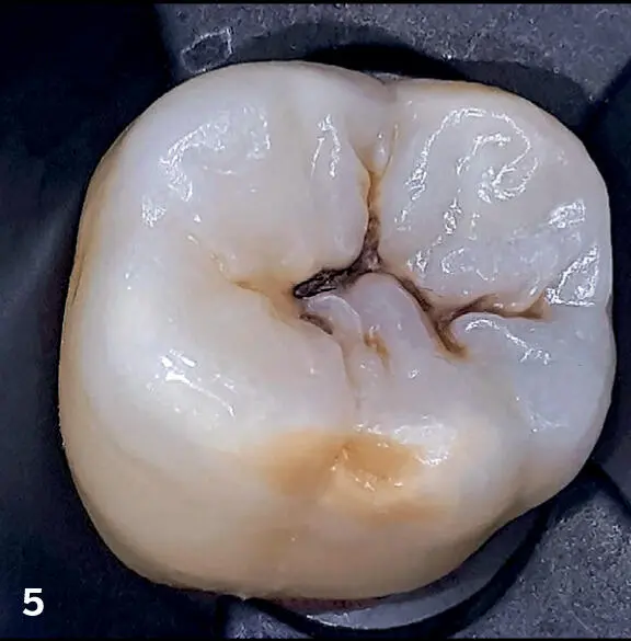
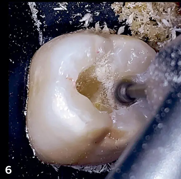
FIGS 5 & 6Patients should know that sometimes a small black line on a tooth can hide a big carious lesion and that they should consult a dentist when they see such a lesion on their teeth. This information can be very empowering for patients who want to be in control of their dental health.
Good communication should also exist between the dentist and the dental technician, especially in the domain of esthetic dentistry. Even the most detailed description written by the dentist cannot compare to the information communicated with a well-shot photograph. Photographs can help the technician visualize how and where his or her work will integrate in its environment. A large amount of information is transferred via photograph, such as the color, shape, alignment, personalization, translucency, opalescence, and halo effect of adjacent teeth.
In addition to other advanced techniques of communicating color information to the laboratory, the following three photographs are very useful:
1.A photograph of a shade guide positioned edge to edge with the natural tooth to show the difference between them (Fig 7).
2.A polarized version of the previous photograph to show the extension of the translucency and its location in the incisal edge, the presence of some details like white spots, and also the difference in color with the artificial shade guide (Fig 8).
3.A photograph showing the secondary and tertiary anatomy because esthetics are not only hue, chroma, and value but also anatomy (Fig 9).
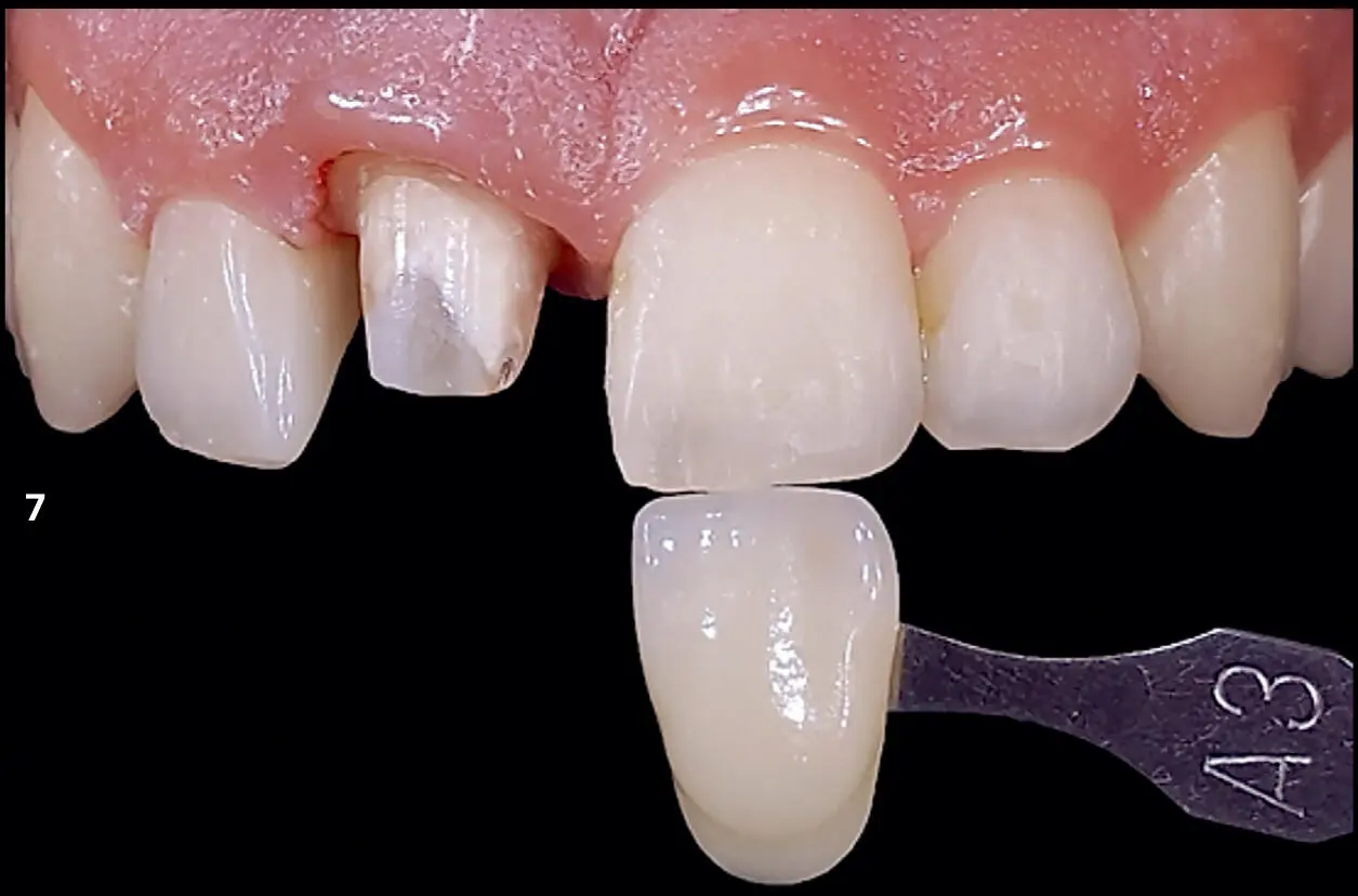
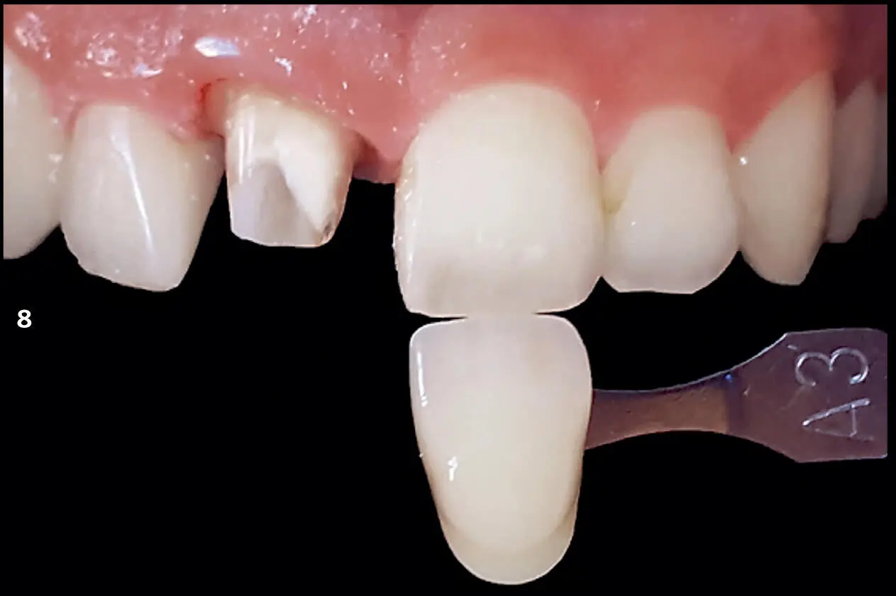
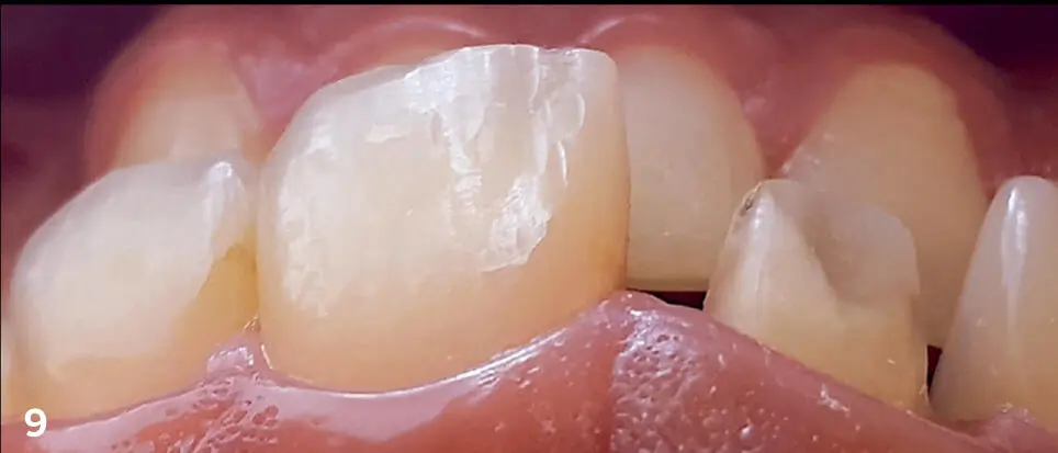
FIGS 7–9The three photographs that should be sent to the dental technician: (1) anterior shot with edge-to-edge shade guide under diffused light; (2) polarized version of edge-to-edge shade guide; (3) shot taken with the light coming from the opposite side of the camera to show anatomical details of the teeth.
Evolution of the Treatment
Photographs are taken prior to any treatment to be used as a baseline indicating the primary situation and to be studied for treatment planning. However, follow-up photographs are just as essential to evaluate the progress of the treatment plan. Photographs can be taken on a regular basis throughout different phases of treatment. Thus, the evolution of the clinical situation and the treatment plan can be monitored according to a certain time interval. This is especially important in specialties like orthodontics, prosthodontics, restorative dentistry, and periodontics. Furthermore, photography allows clinicians to objectively assess any changes in color, shape, or integration of any materials used intraorally (Fig 10), which can aid future treatment planning.
Читать дальшеИнтервал:
Закладка:
Похожие книги на «Protocols for Mobile Dental Photography with Auxiliary Lighting»
Представляем Вашему вниманию похожие книги на «Protocols for Mobile Dental Photography with Auxiliary Lighting» списком для выбора. Мы отобрали схожую по названию и смыслу литературу в надежде предоставить читателям больше вариантов отыскать новые, интересные, ещё непрочитанные произведения.


Обсуждение, отзывы о книге «Protocols for Mobile Dental Photography with Auxiliary Lighting» и просто собственные мнения читателей. Оставьте ваши комментарии, напишите, что Вы думаете о произведении, его смысле или главных героях. Укажите что конкретно понравилось, а что нет, и почему Вы так считаете.