Spectrums of Amyotrophic Lateral Sclerosis
Здесь есть возможность читать онлайн «Spectrums of Amyotrophic Lateral Sclerosis» — ознакомительный отрывок электронной книги совершенно бесплатно, а после прочтения отрывка купить полную версию. В некоторых случаях можно слушать аудио, скачать через торрент в формате fb2 и присутствует краткое содержание. Жанр: unrecognised, на английском языке. Описание произведения, (предисловие) а так же отзывы посетителей доступны на портале библиотеки ЛибКат.
- Название:Spectrums of Amyotrophic Lateral Sclerosis
- Автор:
- Жанр:
- Год:неизвестен
- ISBN:нет данных
- Рейтинг книги:3 / 5. Голосов: 1
-
Избранное:Добавить в избранное
- Отзывы:
-
Ваша оценка:
Spectrums of Amyotrophic Lateral Sclerosis: краткое содержание, описание и аннотация
Предлагаем к чтению аннотацию, описание, краткое содержание или предисловие (зависит от того, что написал сам автор книги «Spectrums of Amyotrophic Lateral Sclerosis»). Если вы не нашли необходимую информацию о книге — напишите в комментариях, мы постараемся отыскать её.
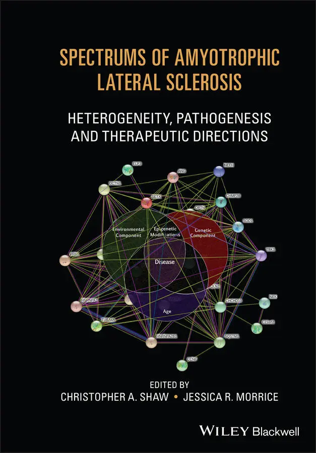
Discover state-of-the-art research findings on ALS from leading authors and editors in the field Spectrums of Amyotrophic Lateral Sclerosis: Heterogeneity, Pathogenesis & Therapeutic Directions
Spectrums of Amyotrophic Lateral Sclerosis: Heterogeneity, Pathogenesis & Therapeutic Directions
Spectrums of Amyotrophic Lateral Sclerosis — читать онлайн ознакомительный отрывок
Ниже представлен текст книги, разбитый по страницам. Система сохранения места последней прочитанной страницы, позволяет с удобством читать онлайн бесплатно книгу «Spectrums of Amyotrophic Lateral Sclerosis», без необходимости каждый раз заново искать на чём Вы остановились. Поставьте закладку, и сможете в любой момент перейти на страницу, на которой закончили чтение.
Интервал:
Закладка:
92 92. Duan, W., Guo, M., Yi, L. et al. (2019). Deletion of Tbk1 disrupts autophagy and reproduces behavioral and locomotor symptoms of FTD‐ALS in mice. Aging (Albany NY) 11 (8): 2457–2476.
93 93. Takahashi, K., Tanabe, K., Ohnuki, M. et al. (2007). Induction of pluripotent stem cells from adult human fibroblasts by defined factors. Cell 131 (5): 861–872.
94 94. Son, E.Y., Ichida, J.K., Wainger, B.J. et al. (2011). Conversion of mouse and human fibroblasts into functional spinal motor neurons. Cell Stem Cell 9 (3): 205–218.
CHAPTER 2 Genetic Basis of ALS
Jay P. Ross 1,2, Patrick A. Dion 2,3, and Guy A. Rouleau 1,2,3
1Department of Human Genetics, McGill University, Montréal, Québec, Canada
2Montreal Neurological Institute and Hospital, McGill University, Montréal, Québec, Canada
3Department of Neurology and Neurosurgery, McGill University, Montréal, Québec, Canada
INTRODUCTION
The recent study of amyotrophic lateral sclerosis (ALS) has generally been driven by its associated genes. While there seems to be a strong environmental component to ALS risk, the heritability of ALS has been estimated to range between 0.523 and 0.61 [1, 2] which suggests a substantial genetic contribution. As the number of cases currently explained by known genetics variants is relatively small, there are likely many genetic contributors yet to be discovered. Further complicating ALS genetics is the distinction between familial and sporadic cases: those with a family history of ALS or frontotemporal dementia, and those without, respectively.
One of the first goals of ALS genetic research has been to identify variants in genes and to fit these genes into our current understanding of ALS biology. Over the years, various methods of gene discovery have been applied. Over time, studies focused on familial linkage, as well as screening of candidate genes encoding a protein with known biological relevance, were replaced by whole‐exome (WES) and whole‐genome sequencing (WGS) studies; such advances have led to the identification of variants more frequently observed in ALS patients than unaffected individuals.
This chapter will review genes for which there is consistent strong evidence and genes that were recently discovered, explore aspects of inheritance for ALS‐associated variants, and consider regulatory and epigenetic variants.
GENES CAUSING ALS
The following four genes are described in order of discovery and are highlighted due to their importance in ALS. Dysfunction and dysregulation of these genes cause ALS, but even for conclusively causal variants in these genes, the most critical disease mechanisms are still under investigation.
Superoxide Dismutase 1 (SOD1)
The first gene in which variants were associated with ALS pathology was superoxide dismutase 1 ( SOD1 ) [3]. First discovered following a polymerase chain reaction (PCR) mutation screening approach of the gene closest to a prior linkage signal, SOD1 variants remained one of the only known genetic causes of ALS for 15 years. Most SOD1 variants lead to a highly penetrant, familial, and dominant presentation of ALS [4], and they are observed in approximately 12% of familial and 1% of sporadic ALS cases [4]. Certain variants (p.D90A, for example) are associated with either dominant or recessive inheritance depending on the population [5, 6]. While SOD1 variants occur in any exon, the frequency of specific SOD1 variants is associated with different ethnicities [5, 7]. For example, SOD1 variants are relatively rare in European cohorts but are the most frequent cause of familial ALS in Asian cohorts [7]. Variant penetrance can be as high as 90–95% by the age of 80 [8], suggesting that the encoded SOD1 protein is intolerant to protein‐altering variants [9] and that most variants are probably associated with familial ALS given sufficient age.
Despite decades of research, the mechanism of SOD1 toxicity in ALS is still not definite. Aggregation of the SOD1 protein is observed with nearly all protein‐altering SOD1 variants [9–11]. Nonetheless, it is uncertain how these SOD1‐positive aggregates are related to the onset and progression of the disease. Variant SOD1 also leads to cell death in the form of excitotoxicity, in which glutamate activity causes prolonged calcium influx and subsequent mitochondrial dysfunction [12]. However, there is evidence that spinal cords with the highest level of SOD1 aggregates are those of longer‐survival ALS [13]. One explanation is that the level of SOD1 aggregates increases in response to normal cellular processes that sequester SOD1 and lower their toxicity. Another potential mechanism for SOD1‐related cell death could be a loss‐of‐function model, in which variants in SOD1 result in a lack of functional SOD1 protein, although such a mechanism has not been convincingly linked to ALS. SOD1 aggregates can recruit wild‐type SOD1 protein [12, 14], leading to a lack of functional SOD1 in the cell. A reduction in SOD1 normal activity, that of a dismutase lowering the number of superoxide radicals, is observed in SOD1 variant carriers [14]. The resulting increase of free radicals in cells has direct negative effects on mitochondrial function and survival. While this observation does not prove that loss of function is the model by which SOD1 variants cause ALS, it does suggest that the mechanism is more complicated than simply SOD1 aggregates causing motor neuron death.
TAR DNA‐Binding Protein 43 (TDP‐43)
Informed by prior observations of transactivation response (TAR) DNA binding protein 43 (TDP‐43) aggregates in neurons of ALS patients, the gene that encodes TDP‐43 ( TARDBP ) was screened for the presence of protein‐altering variants in simultaneous studies [15, 16]. Variants in TARDBP are a rare cause of ALS and are mainly clustered in the C‐terminal low‐complexity domain [17]. TARDBP variants account for 1–4% of familial ALS and are very rare in sporadic ALS [18], suggesting that altered TDP‐43 is a penetrant mechanism for ALS. TARDBP variants generally cause a dominant familial ALS [17] and are observed across ethnicities [7]. Certain founder populations can lead to a significantly increased incidence of TARDBP variants; for example, between 20 and 30% of Sardinian ALS patients carry the p.A382T variant [19, 20]. Despite the rarity of TARDBP variants, the implication of this gene has opened several avenues in ALS research.
TDP‐43 is an RNA binding protein, and it has specific transcript targets, which it binds and modifies [21, 22]. Normal TDP‐43 function involves repressing cryptic exons in mRNA, as variant (and silenced) TARDBP results in exons arising from intronic regions [23–25]. These cryptic exons are generally in nonconserved genomic regions [25]. Transcripts with cryptic exons tend to be processed through nonsense‐mediated decay, and lower expression of normal transcripts therefore results from altered TDP‐43 [24]. A loss‐of‐function model for TDP‐43 implicates not only mis‐spliced targets but also a lower expression of these targets. Conversely, conserved exons that should be included in properly‐spliced transcripts are skipped ( skiptic ) in cells with TDP‐43 variants [26]. In mice carrying certain variants in either the RNA‐binding or low‐complexity domains, exons were either aberrantly included (cryptic) or excluded (skiptic), respectively [26].
Cytoplasmic TDP‐43–containing inclusions are a common hallmark of ALS, appearing in as many as 97% of ALS cases [27] and in all but SOD1 variant carriers [28]. There is still debate about whether the aggregates are toxic per se . First, while most ALS‐causing variants occur in the C‐terminal region of the TARDBP transcript, aggregated C‐terminal TDP‐43 fragments do not appear to be the driving force of the pathology [29]. Second, the N‐terminus of TDP‐43 appears to strongly affect cell pathology and aggregation [30], although ALS‐associated variants are not often observed in the first exons of TARDBP . Lastly, the normal function of the RNA‐binding domains of TDP‐43 appears to prevent the aggregation of the protein: when RNA targets of TDP‐43 are not available to bind, aggregation of TDP‐43 increases [31]. Supporting the involvement of RNA‐binding for ALS pathology, TARDBP variants require intact TDP‐43 RNA‐binding domains to exert neurotoxic effects [32].
Читать дальшеИнтервал:
Закладка:
Похожие книги на «Spectrums of Amyotrophic Lateral Sclerosis»
Представляем Вашему вниманию похожие книги на «Spectrums of Amyotrophic Lateral Sclerosis» списком для выбора. Мы отобрали схожую по названию и смыслу литературу в надежде предоставить читателям больше вариантов отыскать новые, интересные, ещё непрочитанные произведения.
![Ники Сегнит - Тезаурус вкусов 2. Lateral Cooking [litres]](/books/430786/niki-segnit-tezaurus-vkusov-2-lateral-cooking-li-thumb.webp)

Обсуждение, отзывы о книге «Spectrums of Amyotrophic Lateral Sclerosis» и просто собственные мнения читателей. Оставьте ваши комментарии, напишите, что Вы думаете о произведении, его смысле или главных героях. Укажите что конкретно понравилось, а что нет, и почему Вы так считаете.