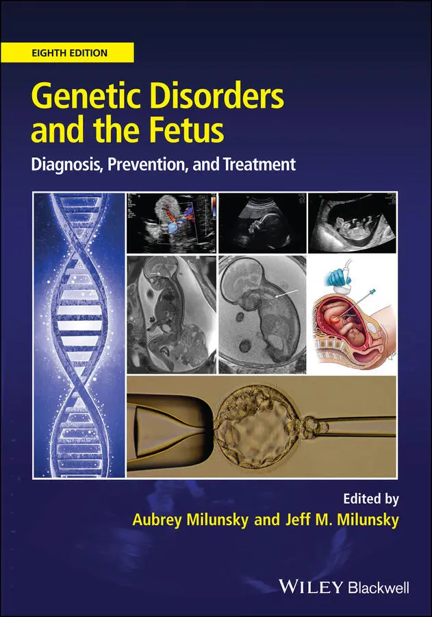
Genetic Disorders and the Fetus
Здесь есть возможность читать онлайн «Genetic Disorders and the Fetus» — ознакомительный отрывок электронной книги совершенно бесплатно, а после прочтения отрывка купить полную версию. В некоторых случаях можно слушать аудио, скачать через торрент в формате fb2 и присутствует краткое содержание. Жанр: unrecognised, на английском языке. Описание произведения, (предисловие) а так же отзывы посетителей доступны на портале библиотеки ЛибКат.
- Название:Genetic Disorders and the Fetus
- Автор:
- Жанр:
- Год:неизвестен
- ISBN:нет данных
- Рейтинг книги:4 / 5. Голосов: 1
-
Избранное:Добавить в избранное
- Отзывы:
-
Ваша оценка:
Genetic Disorders and the Fetus: краткое содержание, описание и аннотация
Предлагаем к чтению аннотацию, описание, краткое содержание или предисловие (зависит от того, что написал сам автор книги «Genetic Disorders and the Fetus»). Если вы не нашли необходимую информацию о книге — напишите в комментариях, мы постараемся отыскать её.
Genetic Disorders and the Fetus — читать онлайн ознакомительный отрывок
Ниже представлен текст книги, разбитый по страницам. Система сохранения места последней прочитанной страницы, позволяет с удобством читать онлайн бесплатно книгу «Genetic Disorders and the Fetus», без необходимости каждый раз заново искать на чём Вы остановились. Поставьте закладку, и сможете в любой момент перейти на страницу, на которой закончили чтение.
Интервал:
Закладка:
10 Chapter 16Figure 16.1 RNA “toxicity” models for fragile X‐associated tremor/ataxia syndr...Figure 16.2 Pedigree of family with FXS, premutation, FXPOI, and FXTAS involve...Figure 16.3 Estimated risk for expansion to the full‐mutation range of a trans...
11 Chapter 17Figure 17.1 Sagittal plane of a fetus with sacral agenesis.Figure 17.2 Demonstrating anencephaly at 11 weeks of gestation in sagittal pla...Figure 17.3 Demonstrating anencephaly at 20 weeks of gestation.Figure 17.4 (a) Transverse section of the fetal head at the level of the septu...Figure 17.5 A fetus with spina bifida in the first trimester at 13 weeks of ge...Figure 17.6 Meckel–Gruber syndrome with the association of multicystic kidneys...Figure 17.7 A fetus with severe ventriculomegaly – hydrocephaly.Figure 17.8 A fetus with holoprosencephaly at 14.6 weeks of gestation.Figure 17.9 (a) Transverse and (b) coronal plane of corpus callosum agenesis a...Figure 17.10 (a) Transverse and (b) sagittal plane of Blake's pouch cyst in a ...Figure 17.11 Transverse plane of a fetus with a Dandy–Walker malformation.Figure 17.12 Fetus with a unilateral cleft lip and/or palate presenting at 19....Figure 17.13 Micrognathia demonstrated in a sagittal plane with the fetus show...Figure 17.14 Macrocystic congenital pulmonary airway malformation visible as a...Figure 17.15 Classic ultrasound finding in left congenital diaphragmatic herni...Figure 17.16 Bilateral pleural effusion on a transverse plane of the fetal tho...Figure 17.17 A fetus with hypoplastic left heart syndrome.Figure 17.18 Demonstrating a ventricular septal defect in transverse plane wit...Figure 17.19 Demonstrating an atrioventricular septal defect with the atrioven...Figure 17.20 Fetus with transposition of the great arteries presenting at 20.4...Figure 17.21 Outflow tract of the overriding aorta with ventricular septal def...Figure 17.22 Large omphalocele at 13 weeks of gestation containing liver.Figure 17.23 Gastroschisis in first trimester at 12.0 weeks of gestation. (a) ...Figure 17.24 (a) Transverse plane showing gastroschisis in the second trimeste...Figure 17.25 Body stalk anomaly at 13 weeks of gestation. (a) Mid‐sagittal pla...Figure 17.26 Body stalk anomaly at 10.3 weeks of gestation. (a, b) Mid‐sagitta...Figure 17.27 Bladder exstrophy with nonvisualization of the urinary bladder.Figure 17.28 The “pouch sign” in a fetus with esophageal atresia.Figure 17.29 “Double bubble” sign in a fetus with duodenal atresia demonstrate...Figure 17.30 Simple abdominal cyst in a female fetus.Figure 17.31 Bilateral hydronephrosis demonstrated in transverse plane (a) and...Figure 17.32 Unilateral multicystic dysplastic kidney demonstrated in transver...Figure 17.33 Polycystic kidney disease presenting two large hyperechogenic kid...Figure 17.34 Unilateral renal agenesis demonstrated in coronal plane.Figure 17.35 A fetus with achondroplasia with frontal bossing (a) and widening...Figure 17.36 Fetus with oesteogenesis imperfecta type II with a hypomineralize...Figure 17.37 Fetus with postaxial polydactyly at 20 weeks of gestation.Figure 17.38 Triploidy of maternal origin with severe asymmetric growth restri...
12 Chapter 18Figure 18.1 Two‐dimensional gray scale and color Doppler echocardiography of f...Figure 18.2 Fetal heart rate, time intervals during cardiac cycle, and conduct...Figure 18.3 (a–c) A case of maternal anti‐SSa/SSb‐induced third‐degree atriove...Figure 18.4 Dextroposition of the fetal heart in left‐sided hydrothorax due to...Figure 18.5 An anomaly of the cardiac axis in tetralogy of Fallot. The axis of...Figure 18.6 (a) A case of hypoplastic left heart (HLH) mitral valve atresia/ao...Figure 18.7 (a) Transverse oblique plane of a fetal thorax with bronchopulmona...Figure 18.8 (a) Left‐sided microcystic congenital cystic adenomatatoid malform...Figure 18.9 (a) Longitudinal section of a fetus presenting with congenital hig...Figure 18.10 Left‐sided diaphragmatic hernia with the fetal stomach and the de...Figure 18.11 Typical image of polyhydramnios and absent stomach bubble in the ...Figure 18.12 (a) Double bubble phenomenon (dilated stomach and duodenum) at 32...Figure 18.13 Transverse section of the fetal abdomen showing a large omphaloce...Figure 18.14 (a) Transverse oblique section of the fetal abdomen at the level ...Figure 18.15 Typical images of lower urinary tract obstruction (LUTO). (a) Bil...Figure 18.16 (a, b) Examples of moderate to severe ventriculomegaly in the sec...Figure 18.17 (a–d) Typical cranial signs in open spina bifida. (a) Banana sign...Figure 18.18 (a, b) In alobar holoprosencephaly there is no interhemispheric f...Figure 18.19 (a) The arrangement of anterior horns and cavum septum pellucidum...Figure 18.20 (a, b) In schizencephaly there is a large holohemispheric cleft o...Figure 18.21 In the case of an absent cavum septum pellucidum there two separa...Figure 18.22 (a, b) Cerebellar findings in the oblique transcerebellar plane: ...
13 Chapter 19Figure 19.1 Axial T2‐weighted images (a, c) and T1‐weighted images (b, d) at 2...Figure 19.2 (a) Axial T2‐weighted images showing corpus callosum agenesis at 2...Figure 19.3 (a, b) Axial T2‐weighted images showing toxoplasmosis at 27 weeks:...Figure 19.4 (a) Axial T2‐weighted images at 32 weeks: cervical cystic lymphang...Figure 19.5 (a) Sagittal T2‐weighted images at 30 weeks: huge sacro‐coccygeal ...
14 Chapter 20Figure 20.1 Ultrasound and radiographic images of hypophosphatasia. The chest ...Figure 20.2 Ultrasound image from 18 weeks gestation shows a normal length sca...Figure 20.3 X‐ray of a 20‐week fetus with thanatophoric dysplasia displaying c...Figure 20.4 Growth curves showing the typical pattern of a fetus with achondro...Figure 20.5 Facial profile of a fetus with achondroplasia at 28 weeks gestatio...Figure 20.6 This fetus with thanatophoric dysplasia type I was noted to have s...Figure 20.7 (a) Normal fetus at 18 weeks of age in profile. There is a smooth ...Figure 20.8 Fetus with hypochondroplasia and normal facial profile.Figure 20.9 A normal looking but short femur in a fetus with hypochondroplasia...Figure 20.10 X‐ray of a fetus with osteogenesis imperfecta (OI) type III shows...Figure 20.11 Ultrasound image showing the head of a fetus with osteogenesis im...Figure 20.12 Lateral view of the leg of a fetus with osteogenesis imperfecta t...Figure 20.13 Beaded ribs in a fetus with osteogenesis imperfecta type III at 3...Figure 20.14 This fetus had molecularly confirmed hypochondrogenesis ( COL2A1 m...Figure 20.15 Ultrasound image of fetus from Figure 20.14. Facial profile shows...Figure 20.16 Ultrasound image showing the second fetus of a nonconsanguineous ...
15 Chapter 22Figure 22.1 Amino acid metabolism and fates.
16 Chapter 23Figure 23.1 Phenotypic spectrum in mucopolysaccharidosis I (MPS I). (a) Severe...
17 Chapter 24Figure 24.1 Schematic diagram showing the four identical chemical reactions of...Figure 24.2 Enzymatic organization of the (a) mitochondrial and (b) peroxisoma...Figure 24.3 Schematic diagram showing the enzymatic steps involved in the mito...
18 Chapter 25Figure 25.1 Lysosomal catabolism of some glycosphingolipids. Source: Milunsky ...
19 Chapter 27Figure 27.1 Prenatal diagnosis of α 0‐thalassemia using gap‐PCR to detect the −...Figure 27.2 Restriction enzyme polymerase chain reaction (RE‐PCR) analysis usi...Figure 27.3 The screening of a DNA sample for seven common Mediterranean mutat...Figure 27.4 Prenatal diagnosis of β‐thalassemia by the allele‐specific priming...Figure 27.5 The diagnosis of the sickle cell anemia genotypes by Dde I digestio...
20 Chapter 29Figure 29.1 Stages of twin‐to‐twin transfusion syndrome (TTTS).Figure 29.2 (a) Donor twin with small bladder and severe oligohydramnios. (b) ...Figure 29.3 (a) Laser being used to ablate the individual arteriovenous connec...Figure 29.4 Twin anemia polycythemia sequence (TAPS). Small, unidirectional, u...Figure 29.5 Prenatal ultrasound image of the placenta in twin anemia polycythe...Figure 29.6 A placenta from a twin anemia polycythemia sequence pregnancy at t...Figure 29.7 Selective fetal growth restriction. (a) Type I Doppler flow is nor...Figure 29.8 Left‐sided fetal congenital diaphragmatic hernia, with right lung ...Figure 29.9 This figure shows diagrammatically the process of placing fetal en...Figure 29.10 Coronal ultrasound image showing the tracheal balloon (between th...Figure 29.11 Ultrasound image showing increased size and echogenicity of the r...Figure 29.12 Ultrasound image showing an enlarged fetal bladder with keyhole s...Figure 29.13 Ultrasound image showing bilateral fetal hydronephrosis. Both kid...Figure 29.14 Ultrasound image showing a decompressed bladder with a double pig...Figure 29.15 (a) Trochar and cannula situated within the fetal bladder after b...Figure 29.16 Large left fetal hydrothorax.Figure 29.17 Texas Children's Fetal Center algorithm for the management of fet...Figure 29.18 The same fetus as in Figure 29.16 after insertion of thoraco‐amni...Figure 29.19 Ultrasound image showing a cross‐sectional view of the chest of a...Figure 29.20 Magnetic resonance imaging shows a predominantly cystic fetal lun...Figure 29.21 Ultrasound image showing an amniotic band constricting the right ...Figure 29.22 Ultrasound image showing a velamentous cord insertion and vasa pr...Figure 29.23 Ultrasound image showing a 6 cm solid mass on fetal surface of pl...Figure 29.24 Ultrasound image showing a lumbosacral myeloschisis starting at L...Figure 29.25 (a) Ultrasound image showing a 22‐week fetus with a large, predom...Figure 29.26 Magnetic resonance imaging of the same fetus details the extent o...Figure 29.27 Postoperative magnetic resonance image of the fetus shown in Figu...Figure 29.28 Images showing the fetal heart following atrial septostomy for hy...Figure 29.29 Figure showing diagrammatically the process employed in our fetos...Figure 29.30 Healed and partially healed relaxing incisions at birth.Figure 29.31 (a) Port sites on the uterus after closure at the initial fetal s...Figure 29.32 (a) Preoperative magnetic resonance image showing hindbrain herni...
Читать дальшеИнтервал:
Закладка:
Похожие книги на «Genetic Disorders and the Fetus»
Представляем Вашему вниманию похожие книги на «Genetic Disorders and the Fetus» списком для выбора. Мы отобрали схожую по названию и смыслу литературу в надежде предоставить читателям больше вариантов отыскать новые, интересные, ещё непрочитанные произведения.


Обсуждение, отзывы о книге «Genetic Disorders and the Fetus» и просто собственные мнения читателей. Оставьте ваши комментарии, напишите, что Вы думаете о произведении, его смысле или главных героях. Укажите что конкретно понравилось, а что нет, и почему Вы так считаете.