Genetic Disorders and the Fetus
Здесь есть возможность читать онлайн «Genetic Disorders and the Fetus» — ознакомительный отрывок электронной книги совершенно бесплатно, а после прочтения отрывка купить полную версию. В некоторых случаях можно слушать аудио, скачать через торрент в формате fb2 и присутствует краткое содержание. Жанр: unrecognised, на английском языке. Описание произведения, (предисловие) а так же отзывы посетителей доступны на портале библиотеки ЛибКат.
- Название:Genetic Disorders and the Fetus
- Автор:
- Жанр:
- Год:неизвестен
- ISBN:нет данных
- Рейтинг книги:4 / 5. Голосов: 1
-
Избранное:Добавить в избранное
- Отзывы:
-
Ваша оценка:
Genetic Disorders and the Fetus: краткое содержание, описание и аннотация
Предлагаем к чтению аннотацию, описание, краткое содержание или предисловие (зависит от того, что написал сам автор книги «Genetic Disorders and the Fetus»). Если вы не нашли необходимую информацию о книге — напишите в комментариях, мы постараемся отыскать её.
Genetic Disorders and the Fetus — читать онлайн ознакомительный отрывок
Ниже представлен текст книги, разбитый по страницам. Система сохранения места последней прочитанной страницы, позволяет с удобством читать онлайн бесплатно книгу «Genetic Disorders and the Fetus», без необходимости каждый раз заново искать на чём Вы остановились. Поставьте закладку, и сможете в любой момент перейти на страницу, на которой закончили чтение.
Интервал:
Закладка:
Women with epilepsy on anticonvulsant medications have an increased risk of having offspring with congenital malformations, noted in one study as 2.7‐fold greater than those without epilepsy. 101A Cochrane Epilepsy Group Registry meta‐analysis of 31 studies of pregnant women on anticonvulsants concluded with increased, but variable RR of congenital malformations of 2.01–5.69, the latter figure being for valproate. 102
There have been reports of an increased risk of congenital malformations following the use of assisted reproductive technology (ART) and negated by other studies. 103A 2018 report using a Centers for Disease Control and Prevention (CDC) database of 11,862,780 livebirths (2011–2013) retrospectively analyzed the 71,050 pregnancies conceived by ART. Infants conceived by ART had an increased risk (77/10,000 vs. 25/10,000), an OR of 2.14. 103The cause(s) of this increase – whether due to the ART or the patients' genetic predisposition – remains to be determined.
Lupo et al. 104in a population‐based registry study of over 10 million children in the United States assessed the association of cancer and congenital malformations. They reported that compared to children without congenital anomalies:
children with chromosomal anomalies (n = 539,567) were 11.6 times more likely to be diagnosed with cancer and
children with nonchromosomal congenital anomalies were 2.5 times more likely to have cancer before 18 years of age.
Congenital malformations and infant morbidity and mortality
The leading cause of infant death in the United States in 2014 was congenital malformations, deformations, and chromosomal abnormalities, accounting for 20.4 percent of 4,748 total infant deaths. 13Survival is clearly dependent on the severity or lethality of the congenital defect. The CDC assessed mortality rates for infants born with trisomy 13 and trisomy 18. The authors identified 5,515 infants born with trisomy 13 and 8,750 born with trisomy 18. The median age at death for both trisomy 13 and trisomy 18 was 10 days. Survival to at least 1 year occurred in 5.6 percent of those born with trisomy 13 or trisomy 18. 105An international registry study (2019) from 18 countries revealed prevalence rates of 0.55 and 1.07 per 10,000 births for trisomies 13 and 18, respectively. Death in the first week of life occurred in 45 percent and 42 percent for trisomy 13 and trisomy 18, respectively. Reported mortality rates were 87 percent and 88 percent at 1 year for each of these trisomies. 106A regional study in the Netherlands noted lethal congenital malformations in 51 percent of stillbirths and 70 percent among those who died during the neonatal period. 107A Scottish study focusing on the survival of 6,153 infants with congenital anomalies up to the age of 5 years noted the following survival rates: chromosomal anomalies (48 percent), NTDs (72 percent), respiratory system anomalies (74 percent), congenital heart disease (75 percent), nervous system anomalies (77 percent) and Down syndrome (84 percent). 108The survival rate among males with congenital defects was 84 percent, compared with 97 percent in those born unaffected. 30Liu et al. 109examined temporal changes in fetal and infant deaths caused by congenital malformations in Canada, England, Wales, and the United States. They concluded that the major factor responsible for the accelerated decline in infant deaths was prenatal diagnosis and elective abortion of fetuses with abnormalities. Given the frequency of Down syndrome, a more detailed discussion follows.
Down syndrome
The availability of prenatal diagnosis and maternal serum screening for chromosomal abnormalities has also affected the birth frequency of Down syndrome. One French study of the impact of prenatal diagnosis over a 21‐year period (1979–1999) in a well‐defined population showed a drop of 80 percent in the birth prevalence of Down syndrome. 110A later report from the Paris Registry of Congenital Anomalies (2001–2005) noted a “fairly stable prevalence of Down syndrome (7.1 per 10,000 livebirths) over time.” 111A Scottish study aimed to assess the impact of prenatal diagnosis on the prevalence of Down syndrome from 1980 to 1996. Both births and pregnancy terminations were included. Pregnancy terminations for Down syndrome rose from 29 percent to about 60 percent. 112In contrast, the prevalence of Down syndrome noted by the Dutch Paediatric Surveillance Unit in 2003 was 16 per 10,000 livebirths, exceeding earlier reports and thought to reflect an older maternal age cohort. 113In the United States, a prevalence rate of 8.27 per 10,000 was reported in 2013 with an estimated 250,700 individuals. 114 , 115In Europe, the 2009–2012 prevalence rate was 10.2 per 100,000 livebirths. 116In Japan, the estimated prevalence rate approximates 22 per 10,000 births. 117Many more babies with Down syndrome are born to women under rather than over 35 years of age. There is some evidence that the risk of having Down syndrome offspring in very young mothers is increased, 118 – 121but not in twin pregnancies. 122
The special problems and associated defects in Down syndrome are well known, as is the increasing life expectancy. Studies from Japan, 123Denmark, 124England, 125Australia, 126and Canada 127highlight the increased life expectancy with Down syndrome. Baird and Sadovnick 127reported a large study of 1,610 individuals with Down syndrome identified in more than 1,500,000 consecutive livebirths in British Columbia from 1908 to 1981. They constructed survival curves and a life table for Down syndrome ( Table 1.2) and for the general population. 128Their estimates show that 44.4 percent and 13.6 percent of liveborn individuals with Down syndrome will survive to 60 and 68 years, respectively, compared with 86.4 percent and 78.4 percent of the general population. In another report, 129the authors have analyzed the causes of death in Down syndrome, highlighting congenital defects and cardiovascular and respiratory illnesses as the most important. A UK population prevalence study noted a median life expectancy of 58 years in 2011. 130
Table 1.2 Life expectancy with Down syndrome, between 1908–1981, to age 68 years.
| Age | Total | Survival at start of age interval (percent) |
|---|---|---|
| 5 | 1,020 | 81.05 |
| 10 | 841 | 78.40 |
| 20 | 497 | 75.34 |
| 30 | 91 | 72.12 |
| 40 | 136 | 69.78 |
| 50 | 57 | 60.68 |
| 55 | 31 | 53.96 |
| 60 | 16 | 44.44 |
| 68 | 1 | 13.57 |
Source: Baird and Sadovnick 1989. 127With permission from John Wiley and Sons.
Additional studies of mortality rates in individuals with Down syndrome revealed that those up to about 35 years of age were little different from others with intellectual disability. Thereafter, however, mortality rates in Down syndrome doubled every 6.4 years, compared with 9.6 years for other intellectually disabled individuals. 129Life tables constructed by these authors indicated a life expectancy of 55 years for a 1‐year‐old patient with Down syndrome and mild/moderate developmental delay and a life expectancy of 43 years for a 1‐year‐old patient with Down syndrome more profoundly affected.
A study from the CDC focused on the death certificates of 17,897 individuals with Down syndrome born between 1983 and 1997. 131These authors reported that the median age at death for those with Down syndrome increased from 25 years in 1983 to 49 years in 1997 ( Figure 1.2).
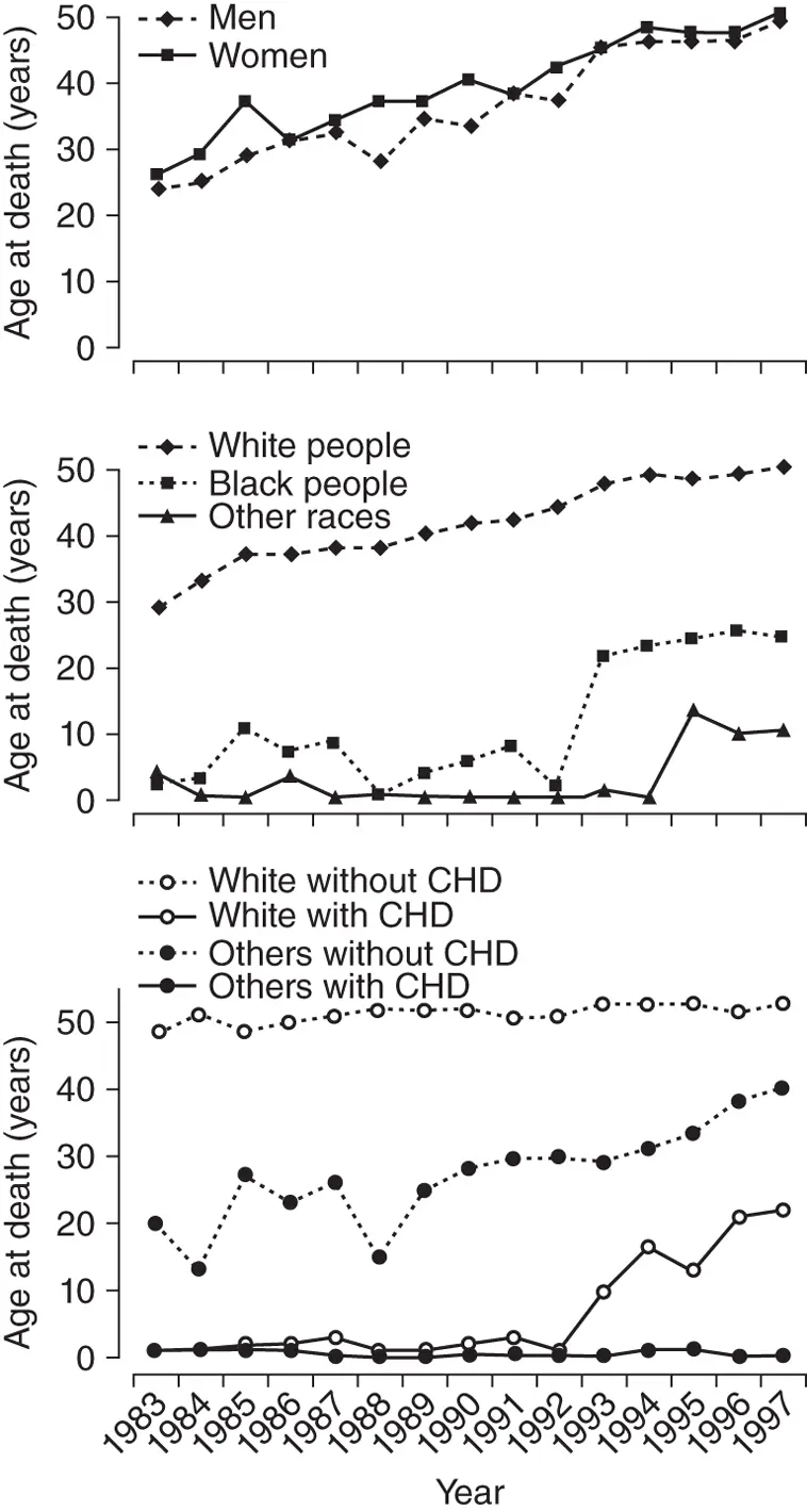
Figure 1.2 Median age at death of people with Down syndrome by sex ( upper ), by racial group ( middle ), and with or without congenital heart defects (CHD) by racial group ( lower ).
Читать дальшеИнтервал:
Закладка:
Похожие книги на «Genetic Disorders and the Fetus»
Представляем Вашему вниманию похожие книги на «Genetic Disorders and the Fetus» списком для выбора. Мы отобрали схожую по названию и смыслу литературу в надежде предоставить читателям больше вариантов отыскать новые, интересные, ещё непрочитанные произведения.


Обсуждение, отзывы о книге «Genetic Disorders and the Fetus» и просто собственные мнения читателей. Оставьте ваши комментарии, напишите, что Вы думаете о произведении, его смысле или главных героях. Укажите что конкретно понравилось, а что нет, и почему Вы так считаете.