Manual of Equine Anesthesia and Analgesia
Здесь есть возможность читать онлайн «Manual of Equine Anesthesia and Analgesia» — ознакомительный отрывок электронной книги совершенно бесплатно, а после прочтения отрывка купить полную версию. В некоторых случаях можно слушать аудио, скачать через торрент в формате fb2 и присутствует краткое содержание. Жанр: unrecognised, на английском языке. Описание произведения, (предисловие) а так же отзывы посетителей доступны на портале библиотеки ЛибКат.
- Название:Manual of Equine Anesthesia and Analgesia
- Автор:
- Жанр:
- Год:неизвестен
- ISBN:нет данных
- Рейтинг книги:3 / 5. Голосов: 1
-
Избранное:Добавить в избранное
- Отзывы:
-
Ваша оценка:
Manual of Equine Anesthesia and Analgesia: краткое содержание, описание и аннотация
Предлагаем к чтению аннотацию, описание, краткое содержание или предисловие (зависит от того, что написал сам автор книги «Manual of Equine Anesthesia and Analgesia»). Если вы не нашли необходимую информацию о книге — напишите в комментариях, мы постараемся отыскать её.
Provides practical, clinically oriented information on anesthetizing equids Uses a bulleted format designed for fast access of key information Offers step-by-step instructions and diagrams of nerve blocks of the limbs, head, and ophthalmic structures Includes new coverage of topics including regulation of extracellular fluid and blood pressure, acid-base disorders, and hemodynamic effects of autonomic drugs
remains a must-have resource for all equine practitioners and veterinary students involved with anesthetizing horses.
Manual of Equine Anesthesia and Analgesia — читать онлайн ознакомительный отрывок
Ниже представлен текст книги, разбитый по страницам. Система сохранения места последней прочитанной страницы, позволяет с удобством читать онлайн бесплатно книгу «Manual of Equine Anesthesia and Analgesia», без необходимости каждый раз заново искать на чём Вы остановились. Поставьте закладку, и сможете в любой момент перейти на страницу, на которой закончили чтение.
Интервал:
Закладка:
Owners might report that the horse previously had a “bad” or “over” reaction to an anesthetic or sedative drug. These concerns should be investigated.
E Fasting
Fasting (~12 hours) was previously advised because of the potential benefits for lung function and the reduced risk of stomach rupture from trauma at induction or recovery.
Many equine hospitals do not fast horses prior to elective surgery. In one study, the PaO2 values during anesthesia were not significantly better in fasted versus non‐fasted horses, and not fasting might reduce the incidence of postanesthetic colic due to changes in gastrointestinal motility.
However, it is generally the case that grain is removed.
Water should be made available up to the time of surgery.
F Medications
It is best to administer all ancillary drugs (e.g. antimicrobials, anti‐inflammatories) prior to sedation. Sodium penicillin can reduce systolic arterial pressure by 8–15 mmHg in anesthetized horses. If antimicrobials are administered during the anesthetic event, this effect can be minimized by administering the drug slowly.
G Jugular catheter
An intravenous (IV) catheter should always be placed prior to anesthesia.
This reduces the likelihood of perivascular injection and provides ready access for further IV anesthetic or emergency drugs.
H Flushing the oral cavity
It is important to flush food debris from the oral cavity, especially if the airway is to be intubated (see Figure 1.1).
I Removal of shoes
Removal of shoes prevents damage to the horse and hospital flooring.However, removal of shoes is not popular with owners. An alternative is to apply bandage material or tape to improve grip and to cover metal points.
Certainly, loose shoes and nails should be removed.
Removal of shoes and metallic debris is necessary when an MRI is to be performed.
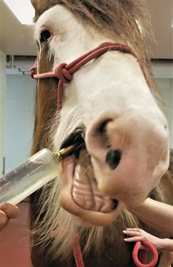
Figure 1.1 Rinsing of the mouth with water prior to induction of anesthesia using a large dosing syringe.
Suggested Reading
1 Bailey, P.A., Hague, B.A., Davis, M. et al. (2016). Incidence of post‐anesthetic colic in non‐fasted adult equine patients. Can. Vet. J. 57: 1263–1266.
2 Dobromylskyj, P., Taylor, P.M., Brearley, J.C. et al. (1996). Effect of pre‐operative starvation on intra‐operative arterial oxygen status in horses. J. Vet. Anaesth. 23: 75–77.
3 Hubbell, J.A.E., Muir, W.W., Robertson, J.T., and Sams, R.A. (1987). Cardiovascular effects of intravenous sodium penicillin, sodium cefazolin, and sodium citrate in awake and anesthetized horses. Vet. Surg. 16: 245–250.
4 Toews, A.R. and Campbell, J.R. (1997). Influence of preoperative complete blood cell counts on surgical outcomes in healthy horses: 102 cases (1986‐1996). J. Am. Vet. Med. Assoc. 211: 887–888.
2 Serum Chemistry and Hematology
Carla Sommardahl
As previously mentioned, there is little value in performing laboratory tests for healthy horses undergoing elective procedures.
However, when indicated, the appropriate tests should, ideally, be performed before general anesthesia is induced. Nevertheless, this may not always be feasible in emergency situations.
I Complete blood count (CBC)
A Erythrocytes
Evaluation of erythrocyte numbers can begin with a determination of the packed cell volume (PCV) or hematocrit (HCT), which measure the percentage of the volume of whole blood that the red blood cells (RBCs) occupy.
Erythrocyte numbers are 6.9–10.7 x 10¹²/l, which represent a normal PCV of 35–45% and a normal hemoglobin of 12–15 g/dl.
A stained blood smear can be used to evaluate RBC morphology and the presence of infectious organisms on the RBCs.
Increased RBC numbers (erythrocytosis) is most commonly associated with hemoconcentration – this is called a relative erythrocytosis as there is no increase in RBC mass.Splenic contraction can cause a transient increase in circulating RBC mass.
In rare cases, there is an increase in numbers due to an increase in RBC production, absolute erythrocytosis.Absolute erythrocytosis is further categorized as primary, as in polycythemia vera, and secondary, as results from chronic hypoxemia.
Decreased RBC numbers (anemia) can be caused by increased removal from the circulation by blood loss or destruction (hemolysis); or by decreased production by the bone marrow.Intravascular hemolysis is accompanied by a decrease in hemoglobin concentration; however, extravascular hemolysis is harder to confirm.Decreased production with iron deficiency secondary to chronic inflammation is a common cause of nonregenerative anemia in the horse, and is characterized by microcytic hypochromic RBCs and decreased RBC indices.Red cell distribution width (RDW) – a measure of RBC variation in size and volume ‐ is used to detect RBC regeneration and is interpreted in conjunction with other values on the complete blood count (CBC).
B Leukocytes
Granulocytes (neutrophils, eosinophils, basophils, and mast cells), monocytes, macrophages, and lymphocytes are important for immune function, and changes in their numbers reflect a response to a disease process.
Total leukocyte count is 5.1–11.0 x 10⁹/l.
Neoplasia, bone marrow disease, or functional defects can cause a decrease or increase in leukocyte numbers.
Neutrophils
Changes in numbers and morphology are important in evaluating the response to systemic inflammation, infection, and stress.
However, the neutrophil count can be normal in the presence of inflammation or infection.
Reference values include 2.8–7.7 x 10⁹/l segmented neutrophils and 0.0–0.2 × 10⁹/l bands.
Neutrophilia is often associated with chronic bacterial infections.
Neutropenia in adult horses is most often associated with endotoxemia (and other bacterial by‐products).
Neutropenia in foals is most commonly associated with sepsis and systemic inflammation.
Eosinophils
Reference values are 0.0–0.7 × 10⁹/l.
Eosinophilia is rare in horses. Conditions to rule out include:Parasitism with cyathastomes.Eosinophilic colitis, enteritis, or multisystemic disease.
Lymphocytes
Reference values are 1.3–4.7 × 10⁹/l.
Lymphopenia: Glucocorticoid release or administration, viral infection, old age, and immunodeficiency in foals (e.g. combined immunodeficiency disease in Arabian or Arabian crossbred foals).
Lymphocytosis: epinephrine release or administration, exercise, equine herpesvirus 2 (foals), leukemia.
Monocytes and Basophils
Reference values for monocytes are 0.1–0.8 × 10⁹/l.
Monocytosis: Chronic inflammation, use of corticosteroids.
Reference values for basophils are 0.0–0.1 × 10⁹/l.
C Platelets
The normal platelet count for horses is 75 000–300 000/μl.
Platelets function in hemostasis, inflammatory responses, immunity, tissue regeneration, and disease pathology.
Thrombocytosis (platelet count >400 000/μl)
Reactive or Secondary:
Age < 3 years, intact males, pyrexia, infectious, or inflammatory conditions.
Platelet count: 400 000–850 000/μl.
Primary or Clonal:
A rare chronic myeloproliferative disorder.
Читать дальшеИнтервал:
Закладка:
Похожие книги на «Manual of Equine Anesthesia and Analgesia»
Представляем Вашему вниманию похожие книги на «Manual of Equine Anesthesia and Analgesia» списком для выбора. Мы отобрали схожую по названию и смыслу литературу в надежде предоставить читателям больше вариантов отыскать новые, интересные, ещё непрочитанные произведения.


Обсуждение, отзывы о книге «Manual of Equine Anesthesia and Analgesia» и просто собственные мнения читателей. Оставьте ваши комментарии, напишите, что Вы думаете о произведении, его смысле или главных героях. Укажите что конкретно понравилось, а что нет, и почему Вы так считаете.