George Herbert Betts - The Mind and Its Education
Здесь есть возможность читать онлайн «George Herbert Betts - The Mind and Its Education» — ознакомительный отрывок электронной книги совершенно бесплатно, а после прочтения отрывка купить полную версию. В некоторых случаях можно слушать аудио, скачать через торрент в формате fb2 и присутствует краткое содержание. Жанр: foreign_edu, pedagogy_book, foreign_psychology, на английском языке. Описание произведения, (предисловие) а так же отзывы посетителей доступны на портале библиотеки ЛибКат.
- Название:The Mind and Its Education
- Автор:
- Жанр:
- Год:неизвестен
- ISBN:нет данных
- Рейтинг книги:3 / 5. Голосов: 1
-
Избранное:Добавить в избранное
- Отзывы:
-
Ваша оценка:
The Mind and Its Education: краткое содержание, описание и аннотация
Предлагаем к чтению аннотацию, описание, краткое содержание или предисловие (зависит от того, что написал сам автор книги «The Mind and Its Education»). Если вы не нашли необходимую информацию о книге — напишите в комментариях, мы постараемся отыскать её.
The Mind and Its Education — читать онлайн ознакомительный отрывок
Ниже представлен текст книги, разбитый по страницам. Система сохранения места последней прочитанной страницы, позволяет с удобством читать онлайн бесплатно книгу «The Mind and Its Education», без необходимости каждый раз заново искать на чём Вы остановились. Поставьте закладку, и сможете в любой момент перейти на страницу, на которой закончили чтение.
Интервал:
Закладка:
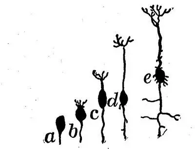
Fig. 6.—Neurones in different stages of development, from a to e . In a , the elementary cell body alone is present; in c , a dendrite is shown projecting upward and an axon downward.—After Donaldson.
The Neurone.—What, then, is a neurone? What is its structure, its function, how does it act? A neurone is a protoplasmic cell, with its outgrowing fibers . The cell part of the neurone is of a variety of shapes, triangular, pyramidal, cylindrical, and irregular. The cells vary in size from 1/250 to 1/3500 of an inch in diameter. In general the function of the cell is thought to be to generate the nervous energy responsible for our consciousness—sensation, memory, reasoning, feeling and all the rest, and for our movements. The cell also provides for the nutrition of the fibers.
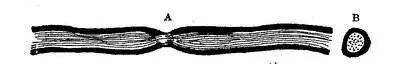
Fig. 7.—Longitudinal (A) and transverse (B) section of nerve fiber. The heavy border represents the medullary, or enveloping sheath, which becomes thicker in the larger fibers.—After Donaldson.
Neurone Fibers.—The neurone fibers are of two kinds, dendrites and axons . The dendrites are comparatively large in diameter, branch freely, like the branches of a tree, and extend but a relatively short distance from the parent cell. Axons are slender, and branch but little, and then approximately at right angles. They reach a much greater distance from the cell body than the dendrites. Neurones vary greatly in length. Some of those found in the spinal cord and brain are not more than 1/12 of an inch long, while others which reach from the extremities to the cord, measure several feet. Both dendrites and axons are of diameter so small as to be invisible except under the microscope.
Neuroglia.—Out of this simple structural element, the neurone, the entire nervous system is built. True, the neurones are held in place, and perhaps insulated, by a kind of soft cement called neuroglia . But this seems to possess no strictly nervous function. The number of the microscopic neurones required to make up the mass of the brain, cord and peripheral nervous system is far beyond our mental grasp. It is computed that the brain and cord contain some 3,000 millions of them.
Complexity of the Brain.—Something of the complexity of the brain structure can best be understood by an illustration. Professor Stratton estimates that if we were to make a model of the human brain, using for the neurone fibers wires so small as to be barely visible to the eye, in order to find room for all the wires the model would need to be the size of a city block on the base and correspondingly high. Imagine a telephone system of this complexity operating from one switch-board!
"Gray" and "White" Matter.—The "gray matter" of the brain and cord is made up of nerve cells and their dendrites, and the terminations of axons, which enter from the adjoining white matter. A part of the mass of gray matter also consists of the neuroglia which surrounds the nerve cells and fibers, and a network of blood vessels. The "white matter" of the central system consists chiefly of axons with their enveloping or medullary, sheath and neuroglia. The white matter contains no nerve cells or dendrites. The difference in color of the gray and the white matter is caused chiefly by the fact that in the gray masses the medullary sheath, which is white, is lacking, thus revealing the ashen gray of the nerve threads. In the white masses the medullary sheath is present.
4. GROSS STRUCTURE OF THE NERVOUS SYSTEM
Divisions of the Nervous System.—The nervous system may be considered in two divisions: (1) The central system, which consists of the brain and spinal cord, and (2) the peripheral system, which comprises the sensory and motor neurones connecting the periphery and the internal organs with the central system and the specialized end-organs of the senses. The sympathetic system, which is found as a double chain of nerve connections joining the roots of sensory and motor nerves just outside the spinal column, does not seem to be directly related to consciousness and so will not be discussed here. A brief description of the nervous system will help us better to understand how its parts all work together in so wonderful a way to accomplish their great result.
The Central System.—In the brain we easily distinguish three major divisions—the cerebrum , the cerebellum and the medulla oblongata . The medulla is but the enlarged upper part of the cord where it connects with the brain. It is about an inch and a quarter long, and is composed of both medullated and unmedullated fibers—that is of both "white" and "gray" matter. In the medulla, the unmedullated neurones which comprise the center of the cord are passing to the outside, and the medullated to the inside, thus taking the positions they occupy in the cerebrum. Here also the neurones are crossing, or changing sides, so that those which pass up the right side of the cord finally connect with the left side of the brain, and vice versa.
The Cerebellum.—Lying just back of the medulla and at the rear part of the base of the cerebrum is the cerebellum, or "little brain," approximately as large as the fist, and composed of a complex arrangement of white and gray matter. Fibers from the spinal cord enter this mass, and others emerge and pass on into the cerebrum, while its two halves also are connected with each other by means of cross fibers.
Fig. 8.—View of the under side of the brain. B, basis of the crura; P, pons; Mo, medulla oblongata; Ce, cerebellum; Sc, spinal cord.
The Cerebrum.—The cerebrum occupies all the upper part of the skull from the front to the rear. It is divided symmetrically into two hemispheres, the right and the left. These hemispheres are connected with each other by a small bridge of fibers called the corpus callosum . Each hemisphere is furrowed and ridged with convolutions, an arrangement which allows greater surface for the distribution of the gray cellular matter over it. Besides these irregularities of surface, each hemisphere is marked also by two deep clefts or fissures —the fissure of Rolando, extending from the middle upper part of the hemisphere downward and forward, passing a little in front of the ear and stopping on a level with the upper part of it; and the fissure of Sylvius, beginning at the base of the brain somewhat in front of the ear and extending upward and backward at an acute angle with the base of the hemisphere.
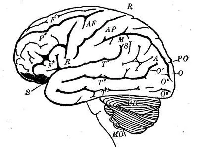
Fig. 9.—Diagrammatic side view of brain, showing cerebellum (CB) and medulla oblongata (MO). F' F'' F''' are placed on the first, second, and third frontal convolutions, respectively; AF, on the ascending frontal; AP, on the ascending parietal; M, on the marginal; A, on the angular. T' T'' T''' are placed on the first, second, and third temporal convolutions. R-R marks the fissure of Rolando; S-S, the fissure of Sylvius; PO, the parieto-occipital fissure.
The surface of each hemisphere may be thought of as mapped out into four lobes: The frontal lobe, which includes the front part of the hemisphere and extends back to the fissure of Rolando and down to the fissure of Sylvius; the parietal lobe, which lies back of the fissure of Rolando and above that of Sylvius and extends back to the occipital lobe; the occipital lobe, which includes the extreme rear portion of the hemisphere; and the temporal lobe, which lies below the fissure of Sylvius and extends back to the occipital lobe.
Читать дальшеИнтервал:
Закладка:
Похожие книги на «The Mind and Its Education»
Представляем Вашему вниманию похожие книги на «The Mind and Its Education» списком для выбора. Мы отобрали схожую по названию и смыслу литературу в надежде предоставить читателям больше вариантов отыскать новые, интересные, ещё непрочитанные произведения.


![Edward Ellis - Adrift on the Pacific - A Boys [sic] Story of the Sea and its Perils](/books/753342/edward-ellis-adrift-on-the-pacific-a-boys-sic-s-thumb.webp)
Обсуждение, отзывы о книге «The Mind and Its Education» и просто собственные мнения читателей. Оставьте ваши комментарии, напишите, что Вы думаете о произведении, его смысле или главных героях. Укажите что конкретно понравилось, а что нет, и почему Вы так считаете.